Calcium »
PDB 1bjq-1byh »
1brw »
Calcium in PDB 1brw: The Crystal Structure of Pyrimidine Nucleoside Phosphorylase in A Closed Conformation
Enzymatic activity of The Crystal Structure of Pyrimidine Nucleoside Phosphorylase in A Closed Conformation
All present enzymatic activity of The Crystal Structure of Pyrimidine Nucleoside Phosphorylase in A Closed Conformation:
2.4.2.2;
2.4.2.2;
Protein crystallography data
The structure of The Crystal Structure of Pyrimidine Nucleoside Phosphorylase in A Closed Conformation, PDB code: 1brw
was solved by
M.J.Pugmire,
S.E.Ealick,
with X-Ray Crystallography technique. A brief refinement statistics is given in the table below:
| Resolution Low / High (Å) | 30.00 / 2.10 |
| Space group | P 1 21 1 |
| Cell size a, b, c (Å), α, β, γ (°) | 53.570, 70.450, 122.780, 90.00, 98.02, 90.00 |
| R / Rfree (%) | 23.2 / 27.6 |
Calcium Binding Sites:
The binding sites of Calcium atom in the The Crystal Structure of Pyrimidine Nucleoside Phosphorylase in A Closed Conformation
(pdb code 1brw). This binding sites where shown within
5.0 Angstroms radius around Calcium atom.
In total 2 binding sites of Calcium where determined in the The Crystal Structure of Pyrimidine Nucleoside Phosphorylase in A Closed Conformation, PDB code: 1brw:
Jump to Calcium binding site number: 1; 2;
In total 2 binding sites of Calcium where determined in the The Crystal Structure of Pyrimidine Nucleoside Phosphorylase in A Closed Conformation, PDB code: 1brw:
Jump to Calcium binding site number: 1; 2;
Calcium binding site 1 out of 2 in 1brw
Go back to
Calcium binding site 1 out
of 2 in the The Crystal Structure of Pyrimidine Nucleoside Phosphorylase in A Closed Conformation
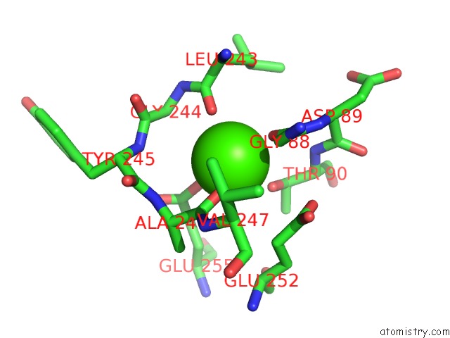
Mono view
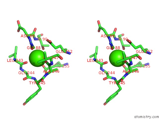
Stereo pair view
Mono view
Stereo pair view
A full contact list of Calcium with other atoms in the Ca binding
site number 1 of The Crystal Structure of Pyrimidine Nucleoside Phosphorylase in A Closed Conformation within 5.0Å range:
|
Calcium binding site 2 out of 2 in 1brw
Go back to
Calcium binding site 2 out
of 2 in the The Crystal Structure of Pyrimidine Nucleoside Phosphorylase in A Closed Conformation
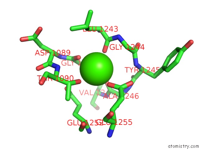
Mono view
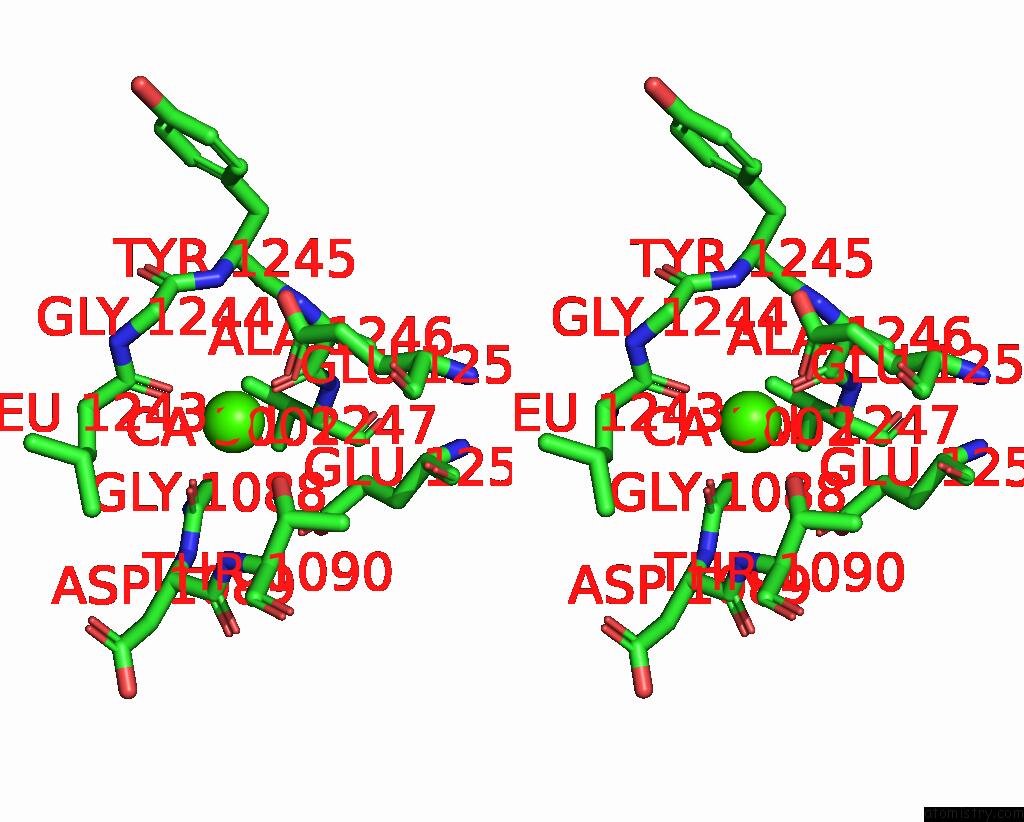
Stereo pair view
Mono view
Stereo pair view
A full contact list of Calcium with other atoms in the Ca binding
site number 2 of The Crystal Structure of Pyrimidine Nucleoside Phosphorylase in A Closed Conformation within 5.0Å range:
|
Reference:
M.J.Pugmire,
S.E.Ealick.
The Crystal Structure of Pyrimidine Nucleoside Phosphorylase in A Closed Conformation. Structure V. 6 1467 1998.
ISSN: ISSN 0969-2126
PubMed: 9817849
DOI: 10.1016/S0969-2126(98)00145-2
Page generated: Mon Jul 7 13:47:37 2025
ISSN: ISSN 0969-2126
PubMed: 9817849
DOI: 10.1016/S0969-2126(98)00145-2
Last articles
Fe in 2YXOFe in 2YRS
Fe in 2YXC
Fe in 2YNM
Fe in 2YVJ
Fe in 2YP1
Fe in 2YU2
Fe in 2YU1
Fe in 2YQB
Fe in 2YOO