Calcium »
PDB 1bjq-1byh »
1byh »
Calcium in PDB 1byh: Molecular and Active-Site Structure of A Bacillus (1-3,1-4)-Beta- Glucanase
Enzymatic activity of Molecular and Active-Site Structure of A Bacillus (1-3,1-4)-Beta- Glucanase
All present enzymatic activity of Molecular and Active-Site Structure of A Bacillus (1-3,1-4)-Beta- Glucanase:
3.2.1.73;
3.2.1.73;
Protein crystallography data
The structure of Molecular and Active-Site Structure of A Bacillus (1-3,1-4)-Beta- Glucanase, PDB code: 1byh
was solved by
T.Keitel,
U.Heinemann,
with X-Ray Crystallography technique. A brief refinement statistics is given in the table below:
| Resolution Low / High (Å) | 8.00 / 2.80 |
| Space group | P 21 21 21 |
| Cell size a, b, c (Å), α, β, γ (°) | 64.320, 78.520, 39.300, 90.00, 90.00, 90.00 |
| R / Rfree (%) | 16.8 / n/a |
Calcium Binding Sites:
The binding sites of Calcium atom in the Molecular and Active-Site Structure of A Bacillus (1-3,1-4)-Beta- Glucanase
(pdb code 1byh). This binding sites where shown within
5.0 Angstroms radius around Calcium atom.
In total only one binding site of Calcium was determined in the Molecular and Active-Site Structure of A Bacillus (1-3,1-4)-Beta- Glucanase, PDB code: 1byh:
In total only one binding site of Calcium was determined in the Molecular and Active-Site Structure of A Bacillus (1-3,1-4)-Beta- Glucanase, PDB code: 1byh:
Calcium binding site 1 out of 1 in 1byh
Go back to
Calcium binding site 1 out
of 1 in the Molecular and Active-Site Structure of A Bacillus (1-3,1-4)-Beta- Glucanase
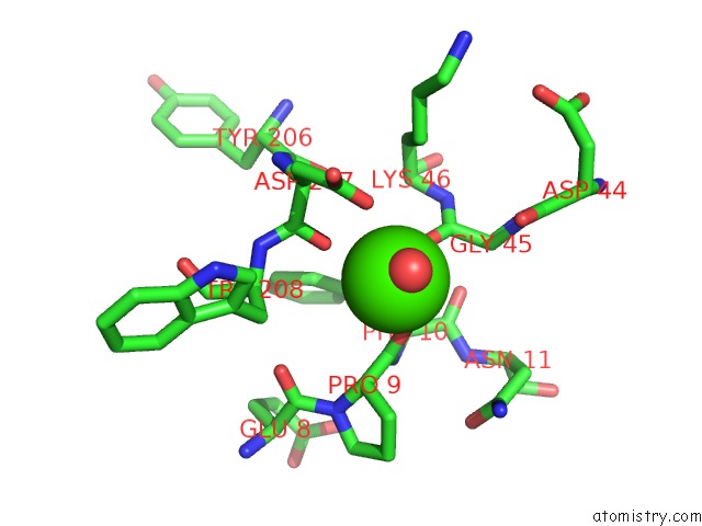
Mono view
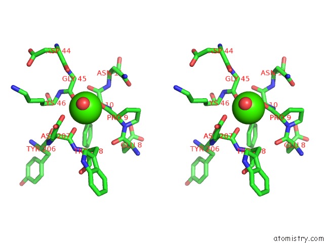
Stereo pair view
Mono view
Stereo pair view
A full contact list of Calcium with other atoms in the Ca binding
site number 1 of Molecular and Active-Site Structure of A Bacillus (1-3,1-4)-Beta- Glucanase within 5.0Å range:
|
Reference:
T.Keitel,
O.Simon,
R.Borriss,
U.Heinemann.
Molecular and Active-Site Structure of A Bacillus 1,3-1,4-Beta-Glucanase. Proc.Natl.Acad.Sci.Usa V. 90 5287 1993.
ISSN: ISSN 0027-8424
PubMed: 8099449
DOI: 10.1073/PNAS.90.11.5287
Page generated: Mon Jul 7 13:49:28 2025
ISSN: ISSN 0027-8424
PubMed: 8099449
DOI: 10.1073/PNAS.90.11.5287
Last articles
Mg in 6TRAMg in 6TR4
Mg in 6TR3
Mg in 6TMF
Mg in 6TQO
Mg in 6TQN
Mg in 6TQF
Mg in 6TQE
Mg in 6TQB
Mg in 6TQA