Calcium »
PDB 1byn-1c5v »
1c0g »
Calcium in PDB 1c0g: Crystal Structure of 1:1 Complex Between Gelsolin Segment 1 and A Dictyostelium/Tetrahymena Chimera Actin (Mutant 228: Q228K/T229A/A230Y/E360H)
Protein crystallography data
The structure of Crystal Structure of 1:1 Complex Between Gelsolin Segment 1 and A Dictyostelium/Tetrahymena Chimera Actin (Mutant 228: Q228K/T229A/A230Y/E360H), PDB code: 1c0g
was solved by
Y.Matsuura,
M.Stewart,
M.Kawamoto,
N.Kamiya,
K.Saeki,
T.Yasunaga,
T.Wakabayashi,
with X-Ray Crystallography technique. A brief refinement statistics is given in the table below:
| Resolution Low / High (Å) | 10.00 / 2.00 |
| Space group | P 21 21 21 |
| Cell size a, b, c (Å), α, β, γ (°) | 56.862, 69.030, 181.500, 90.00, 90.00, 90.00 |
| R / Rfree (%) | 18.2 / 22.3 |
Calcium Binding Sites:
The binding sites of Calcium atom in the Crystal Structure of 1:1 Complex Between Gelsolin Segment 1 and A Dictyostelium/Tetrahymena Chimera Actin (Mutant 228: Q228K/T229A/A230Y/E360H)
(pdb code 1c0g). This binding sites where shown within
5.0 Angstroms radius around Calcium atom.
In total 3 binding sites of Calcium where determined in the Crystal Structure of 1:1 Complex Between Gelsolin Segment 1 and A Dictyostelium/Tetrahymena Chimera Actin (Mutant 228: Q228K/T229A/A230Y/E360H), PDB code: 1c0g:
Jump to Calcium binding site number: 1; 2; 3;
In total 3 binding sites of Calcium where determined in the Crystal Structure of 1:1 Complex Between Gelsolin Segment 1 and A Dictyostelium/Tetrahymena Chimera Actin (Mutant 228: Q228K/T229A/A230Y/E360H), PDB code: 1c0g:
Jump to Calcium binding site number: 1; 2; 3;
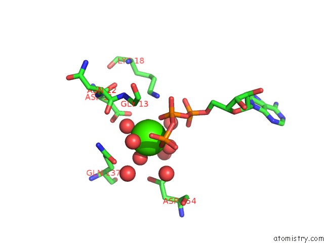
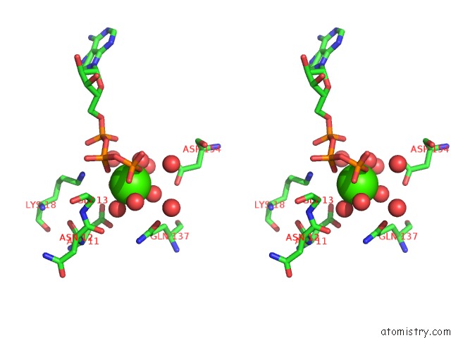
Calcium binding site 1 out of 3 in 1c0g
Go back to
Calcium binding site 1 out
of 3 in the Crystal Structure of 1:1 Complex Between Gelsolin Segment 1 and A Dictyostelium/Tetrahymena Chimera Actin (Mutant 228: Q228K/T229A/A230Y/E360H)
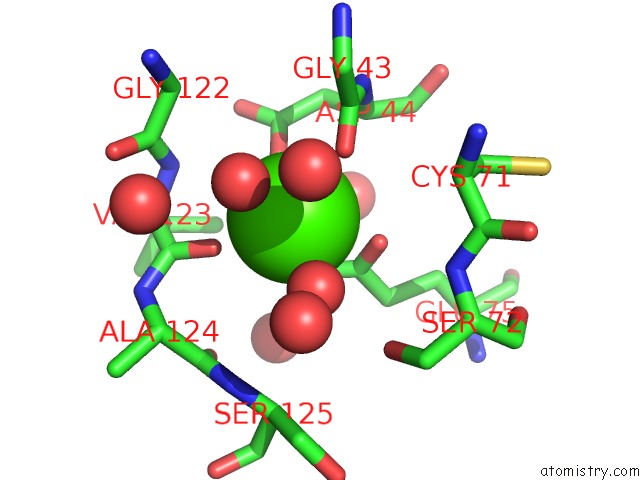
Mono view
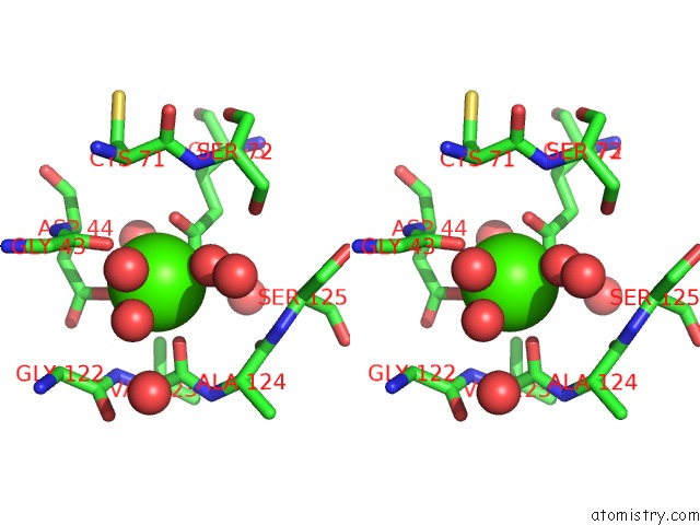
Stereo pair view
Mono view
Stereo pair view
A full contact list of Calcium with other atoms in the Ca binding
site number 1 of Crystal Structure of 1:1 Complex Between Gelsolin Segment 1 and A Dictyostelium/Tetrahymena Chimera Actin (Mutant 228: Q228K/T229A/A230Y/E360H) within 5.0Å range:
|
Calcium binding site 2 out of 3 in 1c0g
Go back to
Calcium binding site 2 out
of 3 in the Crystal Structure of 1:1 Complex Between Gelsolin Segment 1 and A Dictyostelium/Tetrahymena Chimera Actin (Mutant 228: Q228K/T229A/A230Y/E360H)
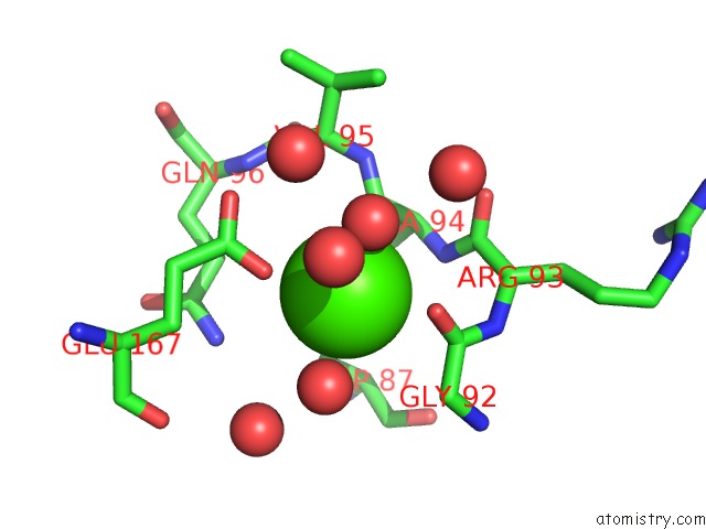
Mono view
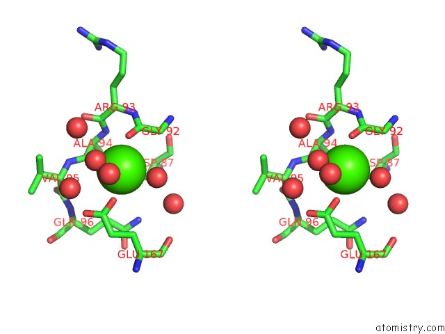
Stereo pair view
Mono view
Stereo pair view
A full contact list of Calcium with other atoms in the Ca binding
site number 2 of Crystal Structure of 1:1 Complex Between Gelsolin Segment 1 and A Dictyostelium/Tetrahymena Chimera Actin (Mutant 228: Q228K/T229A/A230Y/E360H) within 5.0Å range:
|
Calcium binding site 3 out of 3 in 1c0g
Go back to
Calcium binding site 3 out
of 3 in the Crystal Structure of 1:1 Complex Between Gelsolin Segment 1 and A Dictyostelium/Tetrahymena Chimera Actin (Mutant 228: Q228K/T229A/A230Y/E360H)
Mono view
Stereo pair view
Mono view
Stereo pair view
A full contact list of Calcium with other atoms in the Ca binding
site number 3 of Crystal Structure of 1:1 Complex Between Gelsolin Segment 1 and A Dictyostelium/Tetrahymena Chimera Actin (Mutant 228: Q228K/T229A/A230Y/E360H) within 5.0Å range:
|
Reference:
Y.Matsuura,
M.Stewart,
M.Kawamoto,
N.Kamiya,
K.Saeki,
T.Yasunaga,
T.Wakabayashi.
Structural Basis For the Higher Ca(2+)-Activation of the Regulated Actin-Activated Myosin Atpase Observed with Dictyostelium/Tetrahymena Actin Chimeras. J.Mol.Biol. V. 296 579 2000.
ISSN: ISSN 0022-2836
PubMed: 10669610
DOI: 10.1006/JMBI.1999.3467
Page generated: Mon Jul 7 13:50:45 2025
ISSN: ISSN 0022-2836
PubMed: 10669610
DOI: 10.1006/JMBI.1999.3467
Last articles
Mg in 4NXIMg in 4NX8
Mg in 4NV0
Mg in 4NWI
Mg in 4NX5
Mg in 4NV3
Mg in 4NW7
Mg in 4NRU
Mg in 4NST
Mg in 4NUA