Calcium »
PDB 1ck6-1cxi »
1cpn »
Calcium in PDB 1cpn: Native-Like in Vivo Folding of A Circularly Permuted Jellyroll Protein Shown By Crystal Structure Analysis
Enzymatic activity of Native-Like in Vivo Folding of A Circularly Permuted Jellyroll Protein Shown By Crystal Structure Analysis
All present enzymatic activity of Native-Like in Vivo Folding of A Circularly Permuted Jellyroll Protein Shown By Crystal Structure Analysis:
3.2.1.73;
3.2.1.73;
Protein crystallography data
The structure of Native-Like in Vivo Folding of A Circularly Permuted Jellyroll Protein Shown By Crystal Structure Analysis, PDB code: 1cpn
was solved by
M.Hahn,
U.Heinemann,
with X-Ray Crystallography technique. A brief refinement statistics is given in the table below:
| Resolution Low / High (Å) | 8.00 / 1.80 |
| Space group | P 1 21 1 |
| Cell size a, b, c (Å), α, β, γ (°) | 65.470, 41.560, 39.570, 90.00, 111.46, 90.00 |
| R / Rfree (%) | n/a / n/a |
Calcium Binding Sites:
The binding sites of Calcium atom in the Native-Like in Vivo Folding of A Circularly Permuted Jellyroll Protein Shown By Crystal Structure Analysis
(pdb code 1cpn). This binding sites where shown within
5.0 Angstroms radius around Calcium atom.
In total only one binding site of Calcium was determined in the Native-Like in Vivo Folding of A Circularly Permuted Jellyroll Protein Shown By Crystal Structure Analysis, PDB code: 1cpn:
In total only one binding site of Calcium was determined in the Native-Like in Vivo Folding of A Circularly Permuted Jellyroll Protein Shown By Crystal Structure Analysis, PDB code: 1cpn:
Calcium binding site 1 out of 1 in 1cpn
Go back to
Calcium binding site 1 out
of 1 in the Native-Like in Vivo Folding of A Circularly Permuted Jellyroll Protein Shown By Crystal Structure Analysis
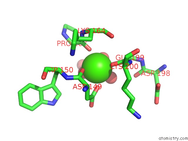
Mono view
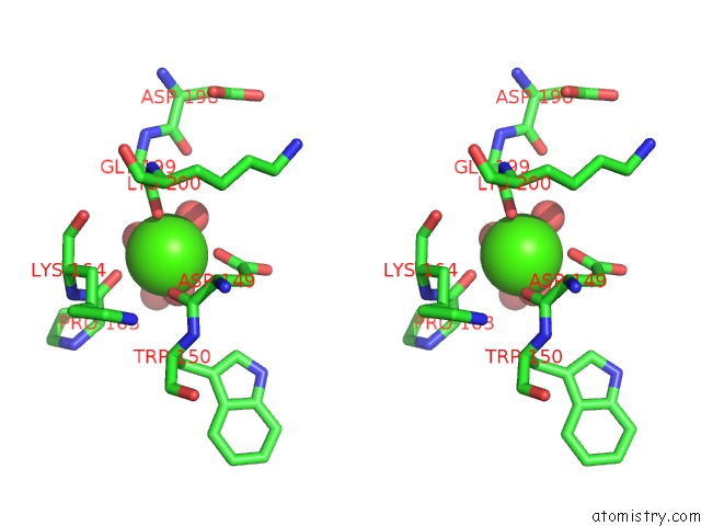
Stereo pair view
Mono view
Stereo pair view
A full contact list of Calcium with other atoms in the Ca binding
site number 1 of Native-Like in Vivo Folding of A Circularly Permuted Jellyroll Protein Shown By Crystal Structure Analysis within 5.0Å range:
|
Reference:
M.Hahn,
K.Piotukh,
R.Borriss,
U.Heinemann.
Native-Like in Vivo Folding of A Circularly Permuted Jellyroll Protein Shown By Crystal Structure Analysis. Proc.Natl.Acad.Sci.Usa V. 91 10417 1994.
ISSN: ISSN 0027-8424
PubMed: 7937966
DOI: 10.1073/PNAS.91.22.10417
Page generated: Mon Jul 7 14:06:41 2025
ISSN: ISSN 0027-8424
PubMed: 7937966
DOI: 10.1073/PNAS.91.22.10417
Last articles
Pd in 3ESDPd in 3AF9
Pd in 3C6S
Pd in 3BZ4
Pd in 3AF8
Pd in 2ZG8
Pd in 2ZG9
Pd in 2Z5Q
Pd in 2Z5R
Pd in 2ZG7