Calcium »
PDB 1de4-1dv8 »
1dpo »
Calcium in PDB 1dpo: Structure of Rat Trypsin
Enzymatic activity of Structure of Rat Trypsin
All present enzymatic activity of Structure of Rat Trypsin:
3.4.21.4;
3.4.21.4;
Protein crystallography data
The structure of Structure of Rat Trypsin, PDB code: 1dpo
was solved by
R.M.Stroud,
with X-Ray Crystallography technique. A brief refinement statistics is given in the table below:
| Resolution Low / High (Å) | 12.00 / 1.59 |
| Space group | I 2 3 |
| Cell size a, b, c (Å), α, β, γ (°) | 123.120, 123.120, 123.120, 90.00, 90.00, 90.00 |
| R / Rfree (%) | 17.4 / n/a |
Calcium Binding Sites:
The binding sites of Calcium atom in the Structure of Rat Trypsin
(pdb code 1dpo). This binding sites where shown within
5.0 Angstroms radius around Calcium atom.
In total only one binding site of Calcium was determined in the Structure of Rat Trypsin, PDB code: 1dpo:
In total only one binding site of Calcium was determined in the Structure of Rat Trypsin, PDB code: 1dpo:
Calcium binding site 1 out of 1 in 1dpo
Go back to
Calcium binding site 1 out
of 1 in the Structure of Rat Trypsin
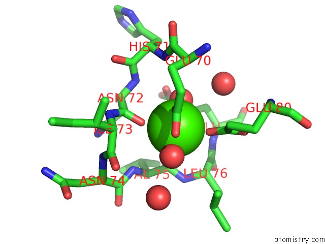
Mono view
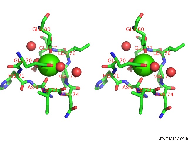
Stereo pair view
Mono view
Stereo pair view
A full contact list of Calcium with other atoms in the Ca binding
site number 1 of Structure of Rat Trypsin within 5.0Å range:
|
Reference:
T.Earnest,
E.Fauman,
C.S.Craik,
R.Stroud.
1.59 A Structure of Trypsin at 120 K: Comparison of Low Temperature and Room Temperature Structures. Proteins V. 10 171 1991.
ISSN: ISSN 0887-3585
PubMed: 1881877
DOI: 10.1002/PROT.340100303
Page generated: Mon Jul 7 14:28:17 2025
ISSN: ISSN 0887-3585
PubMed: 1881877
DOI: 10.1002/PROT.340100303
Last articles
Fe in 2YXOFe in 2YRS
Fe in 2YXC
Fe in 2YNM
Fe in 2YVJ
Fe in 2YP1
Fe in 2YU2
Fe in 2YU1
Fe in 2YQB
Fe in 2YOO