Calcium »
PDB 1de4-1dv8 »
1drb »
Calcium in PDB 1drb: Crystal Structure of Unliganded Escherichia Coli Dihydrofolate Reductase. Ligand-Induced Conformational Changes and Cooperativity in Binding
Enzymatic activity of Crystal Structure of Unliganded Escherichia Coli Dihydrofolate Reductase. Ligand-Induced Conformational Changes and Cooperativity in Binding
All present enzymatic activity of Crystal Structure of Unliganded Escherichia Coli Dihydrofolate Reductase. Ligand-Induced Conformational Changes and Cooperativity in Binding:
1.5.1.3;
1.5.1.3;
Protein crystallography data
The structure of Crystal Structure of Unliganded Escherichia Coli Dihydrofolate Reductase. Ligand-Induced Conformational Changes and Cooperativity in Binding, PDB code: 1drb
was solved by
C.David,
J.Kraut,
with X-Ray Crystallography technique. A brief refinement statistics is given in the table below:
| Resolution Low / High (Å) | 5.00 / 1.96 |
| Space group | P 61 |
| Cell size a, b, c (Å), α, β, γ (°) | 92.840, 92.840, 74.240, 90.00, 90.00, 120.00 |
| R / Rfree (%) | n/a / n/a |
Other elements in 1drb:
The structure of Crystal Structure of Unliganded Escherichia Coli Dihydrofolate Reductase. Ligand-Induced Conformational Changes and Cooperativity in Binding also contains other interesting chemical elements:
| Chlorine | (Cl) | 2 atoms |
Calcium Binding Sites:
The binding sites of Calcium atom in the Crystal Structure of Unliganded Escherichia Coli Dihydrofolate Reductase. Ligand-Induced Conformational Changes and Cooperativity in Binding
(pdb code 1drb). This binding sites where shown within
5.0 Angstroms radius around Calcium atom.
In total only one binding site of Calcium was determined in the Crystal Structure of Unliganded Escherichia Coli Dihydrofolate Reductase. Ligand-Induced Conformational Changes and Cooperativity in Binding, PDB code: 1drb:
In total only one binding site of Calcium was determined in the Crystal Structure of Unliganded Escherichia Coli Dihydrofolate Reductase. Ligand-Induced Conformational Changes and Cooperativity in Binding, PDB code: 1drb:
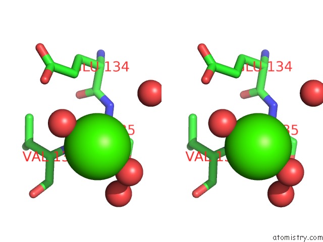
Calcium binding site 1 out of 1 in 1drb
Go back to
Calcium binding site 1 out
of 1 in the Crystal Structure of Unliganded Escherichia Coli Dihydrofolate Reductase. Ligand-Induced Conformational Changes and Cooperativity in Binding

Mono view
Stereo pair view
Mono view
Stereo pair view
A full contact list of Calcium with other atoms in the Ca binding
site number 1 of Crystal Structure of Unliganded Escherichia Coli Dihydrofolate Reductase. Ligand-Induced Conformational Changes and Cooperativity in Binding within 5.0Å range:
|
Reference:
C.Bystroff,
J.Kraut.
Crystal Structure of Unliganded Escherichia Coli Dihydrofolate Reductase. Ligand-Induced Conformational Changes and Cooperativity in Binding. Biochemistry V. 30 2227 1991.
ISSN: ISSN 0006-2960
PubMed: 1998681
DOI: 10.1021/BI00222A028
Page generated: Mon Jul 7 14:29:07 2025
ISSN: ISSN 0006-2960
PubMed: 1998681
DOI: 10.1021/BI00222A028
Last articles
Mg in 4NPWMg in 4NO6
Mg in 4NPV
Mg in 4NOB
Mg in 4NNW
Mg in 4NO1
Mg in 4NNN
Mg in 4NO4
Mg in 4NNB
Mg in 4NMN