Calcium »
PDB 1fak-1fn6 »
1flg »
Calcium in PDB 1flg: Crystal Structure of the Quinoprotein Ethanol Dehydrogenase From Pseudomonas Aeruginosa
Protein crystallography data
The structure of Crystal Structure of the Quinoprotein Ethanol Dehydrogenase From Pseudomonas Aeruginosa, PDB code: 1flg
was solved by
T.Keitel,
A.Diehl,
T.Knaute,
J.J.Stezowski,
W.Hohne,
H.Gorisch,
with X-Ray Crystallography technique. A brief refinement statistics is given in the table below:
| Resolution Low / High (Å) | 12.50 / 2.60 |
| Space group | H 3 |
| Cell size a, b, c (Å), α, β, γ (°) | 159.400, 159.400, 130.950, 90.00, 90.00, 120.00 |
| R / Rfree (%) | 19.2 / 27.4 |
Calcium Binding Sites:
The binding sites of Calcium atom in the Crystal Structure of the Quinoprotein Ethanol Dehydrogenase From Pseudomonas Aeruginosa
(pdb code 1flg). This binding sites where shown within
5.0 Angstroms radius around Calcium atom.
In total 4 binding sites of Calcium where determined in the Crystal Structure of the Quinoprotein Ethanol Dehydrogenase From Pseudomonas Aeruginosa, PDB code: 1flg:
Jump to Calcium binding site number: 1; 2; 3; 4;
In total 4 binding sites of Calcium where determined in the Crystal Structure of the Quinoprotein Ethanol Dehydrogenase From Pseudomonas Aeruginosa, PDB code: 1flg:
Jump to Calcium binding site number: 1; 2; 3; 4;
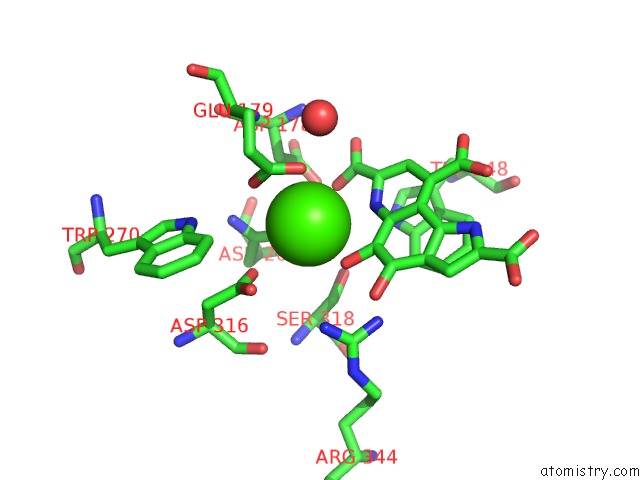
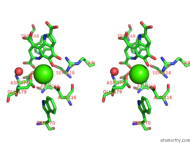
Calcium binding site 1 out of 4 in 1flg
Go back to
Calcium binding site 1 out
of 4 in the Crystal Structure of the Quinoprotein Ethanol Dehydrogenase From Pseudomonas Aeruginosa
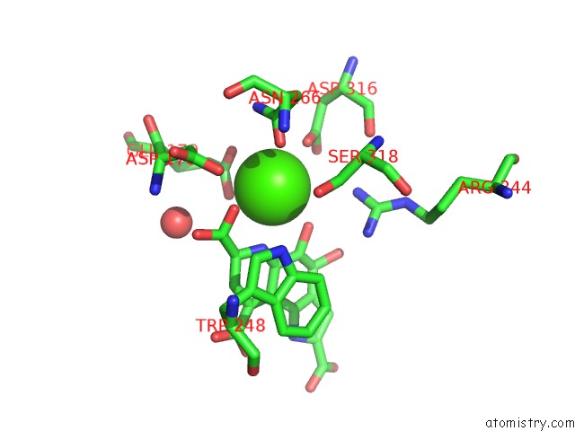
Mono view
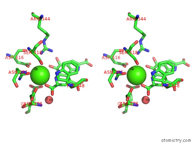
Stereo pair view
Mono view
Stereo pair view
A full contact list of Calcium with other atoms in the Ca binding
site number 1 of Crystal Structure of the Quinoprotein Ethanol Dehydrogenase From Pseudomonas Aeruginosa within 5.0Å range:
|
Calcium binding site 2 out of 4 in 1flg
Go back to
Calcium binding site 2 out
of 4 in the Crystal Structure of the Quinoprotein Ethanol Dehydrogenase From Pseudomonas Aeruginosa
Mono view
Stereo pair view
Mono view
Stereo pair view
A full contact list of Calcium with other atoms in the Ca binding
site number 2 of Crystal Structure of the Quinoprotein Ethanol Dehydrogenase From Pseudomonas Aeruginosa within 5.0Å range:
|
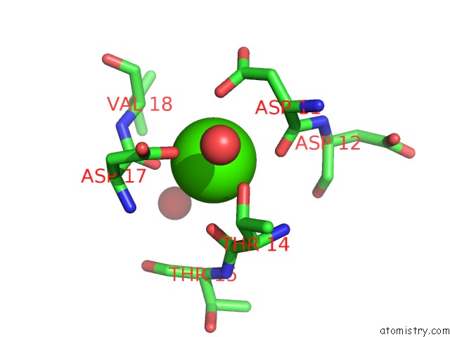
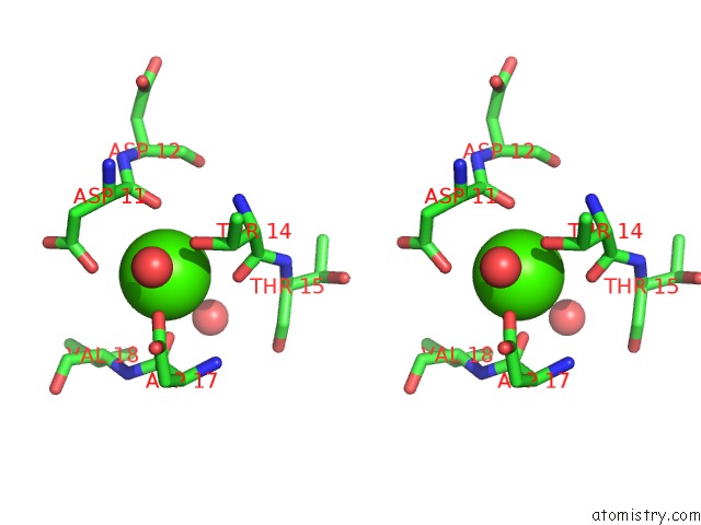
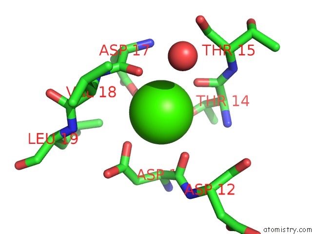
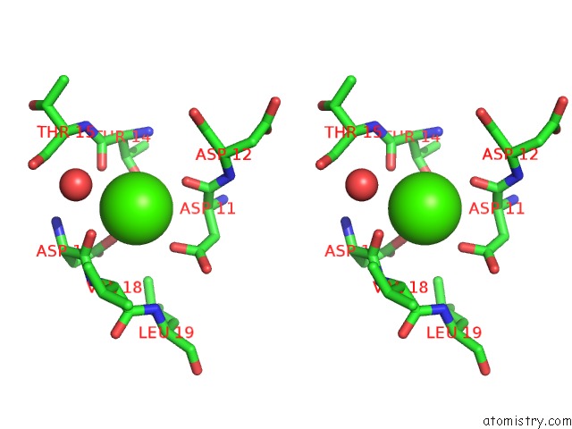
Calcium binding site 3 out of 4 in 1flg
Go back to
Calcium binding site 3 out
of 4 in the Crystal Structure of the Quinoprotein Ethanol Dehydrogenase From Pseudomonas Aeruginosa
Mono view
Stereo pair view
Mono view
Stereo pair view
A full contact list of Calcium with other atoms in the Ca binding
site number 3 of Crystal Structure of the Quinoprotein Ethanol Dehydrogenase From Pseudomonas Aeruginosa within 5.0Å range:
|
Calcium binding site 4 out of 4 in 1flg
Go back to
Calcium binding site 4 out
of 4 in the Crystal Structure of the Quinoprotein Ethanol Dehydrogenase From Pseudomonas Aeruginosa
Mono view
Stereo pair view
Mono view
Stereo pair view
A full contact list of Calcium with other atoms in the Ca binding
site number 4 of Crystal Structure of the Quinoprotein Ethanol Dehydrogenase From Pseudomonas Aeruginosa within 5.0Å range:
|
Reference:
T.Keitel,
A.Diehl,
T.Knaute,
J.J.Stezowski,
W.Hohne,
H.Gorisch.
X-Ray Structure of the Quinoprotein Ethanol Dehydrogenase From Pseudomonas Aeruginosa: Basis of Substrate Specificity. J.Mol.Biol. V. 297 961 2000.
ISSN: ISSN 0022-2836
PubMed: 10736230
DOI: 10.1006/JMBI.2000.3603
Page generated: Mon Jul 7 14:55:05 2025
ISSN: ISSN 0022-2836
PubMed: 10736230
DOI: 10.1006/JMBI.2000.3603
Last articles
Mg in 4B2QMg in 4B3A
Mg in 4B1Z
Mg in 4B2P
Mg in 4B2M
Mg in 4B2K
Mg in 4B2J
Mg in 4B2D
Mg in 4B20
Mg in 4B2H