Calcium »
PDB 1fak-1fn6 »
1fml »
Calcium in PDB 1fml: Crystal Structure of Retinol Dehydratase in A Complex with Retinol and Pap
Protein crystallography data
The structure of Crystal Structure of Retinol Dehydratase in A Complex with Retinol and Pap, PDB code: 1fml
was solved by
S.Pakhomova,
M.Kobayashi,
J.Buck,
M.E.Newcomer,
with X-Ray Crystallography technique. A brief refinement statistics is given in the table below:
| Resolution Low / High (Å) | 38.45 / 2.75 |
| Space group | P 1 21 1 |
| Cell size a, b, c (Å), α, β, γ (°) | 82.053, 66.612, 84.904, 90.00, 111.29, 90.00 |
| R / Rfree (%) | 22.2 / 27.3 |
Calcium Binding Sites:
The binding sites of Calcium atom in the Crystal Structure of Retinol Dehydratase in A Complex with Retinol and Pap
(pdb code 1fml). This binding sites where shown within
5.0 Angstroms radius around Calcium atom.
In total 2 binding sites of Calcium where determined in the Crystal Structure of Retinol Dehydratase in A Complex with Retinol and Pap, PDB code: 1fml:
Jump to Calcium binding site number: 1; 2;
In total 2 binding sites of Calcium where determined in the Crystal Structure of Retinol Dehydratase in A Complex with Retinol and Pap, PDB code: 1fml:
Jump to Calcium binding site number: 1; 2;
Calcium binding site 1 out of 2 in 1fml
Go back to
Calcium binding site 1 out
of 2 in the Crystal Structure of Retinol Dehydratase in A Complex with Retinol and Pap

Mono view
Stereo pair view
Mono view
Stereo pair view
A full contact list of Calcium with other atoms in the Ca binding
site number 1 of Crystal Structure of Retinol Dehydratase in A Complex with Retinol and Pap within 5.0Å range:
|
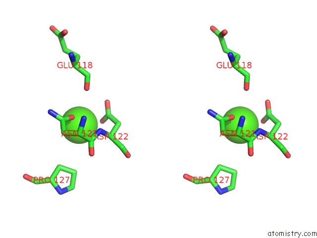
Calcium binding site 2 out of 2 in 1fml
Go back to
Calcium binding site 2 out
of 2 in the Crystal Structure of Retinol Dehydratase in A Complex with Retinol and Pap

Mono view

Stereo pair view
Mono view
Stereo pair view
A full contact list of Calcium with other atoms in the Ca binding
site number 2 of Crystal Structure of Retinol Dehydratase in A Complex with Retinol and Pap within 5.0Å range:
|
Reference:
S.Pakhomova,
M.Kobayashi,
J.Buck,
M.E.Newcomer.
A Helical Lid Converts A Sulfotransferase to A Dehydratase. Nat.Struct.Biol. V. 8 447 2001.
ISSN: ISSN 1072-8368
PubMed: 11323722
DOI: 10.1038/87617
Page generated: Mon Jul 7 14:55:47 2025
ISSN: ISSN 1072-8368
PubMed: 11323722
DOI: 10.1038/87617
Last articles
Mg in 4DUZMg in 4DUY
Mg in 4DR7
Mg in 4DR6
Mg in 4DR5
Mg in 4DUX
Mg in 4DUW
Mg in 4DUV
Mg in 4DUO
Mg in 4DUG