Calcium »
PDB 1fn7-1fzc »
1fxh »
Calcium in PDB 1fxh: Mutant of Penicillin Acylase Impaired in Catalysis with Phenylacetic Acid in the Active Site
Enzymatic activity of Mutant of Penicillin Acylase Impaired in Catalysis with Phenylacetic Acid in the Active Site
All present enzymatic activity of Mutant of Penicillin Acylase Impaired in Catalysis with Phenylacetic Acid in the Active Site:
3.5.1.11;
3.5.1.11;
Protein crystallography data
The structure of Mutant of Penicillin Acylase Impaired in Catalysis with Phenylacetic Acid in the Active Site, PDB code: 1fxh
was solved by
W.B.Alkema,
C.M.Hensgens,
E.H.Kroezinga,
E.De Vries,
R.Floris,
J.M.Van Der Laan,
B.W.Dijkstra,
D.B.Janssen,
with X-Ray Crystallography technique. A brief refinement statistics is given in the table below:
| Resolution Low / High (Å) | 20.00 / 1.97 |
| Space group | P 1 |
| Cell size a, b, c (Å), α, β, γ (°) | 51.014, 64.084, 64.225, 72.92, 73.91, 73.54 |
| R / Rfree (%) | 18.1 / 21.5 |
Calcium Binding Sites:
The binding sites of Calcium atom in the Mutant of Penicillin Acylase Impaired in Catalysis with Phenylacetic Acid in the Active Site
(pdb code 1fxh). This binding sites where shown within
5.0 Angstroms radius around Calcium atom.
In total only one binding site of Calcium was determined in the Mutant of Penicillin Acylase Impaired in Catalysis with Phenylacetic Acid in the Active Site, PDB code: 1fxh:
In total only one binding site of Calcium was determined in the Mutant of Penicillin Acylase Impaired in Catalysis with Phenylacetic Acid in the Active Site, PDB code: 1fxh:
Calcium binding site 1 out of 1 in 1fxh
Go back to
Calcium binding site 1 out
of 1 in the Mutant of Penicillin Acylase Impaired in Catalysis with Phenylacetic Acid in the Active Site
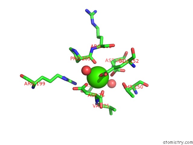
Mono view
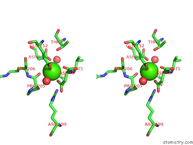
Stereo pair view
Mono view
Stereo pair view
A full contact list of Calcium with other atoms in the Ca binding
site number 1 of Mutant of Penicillin Acylase Impaired in Catalysis with Phenylacetic Acid in the Active Site within 5.0Å range:
|
Reference:
W.B.Alkema,
C.M.Hensgens,
E.H.Kroezinga,
E.De Vries,
R.Floris,
J.M.Van Der Laan,
B.W.Dijkstra,
D.B.Janssen.
Characterization of the Beta-Lactam Binding Site of Penicillin Acylase of Escherichia Coli By Structural and Site-Directed Mutagenesis Studies. Protein Eng. V. 13 857 2000.
ISSN: ISSN 0269-2139
PubMed: 11239085
DOI: 10.1093/PROTEIN/13.12.857
Page generated: Mon Jul 7 14:57:54 2025
ISSN: ISSN 0269-2139
PubMed: 11239085
DOI: 10.1093/PROTEIN/13.12.857
Last articles
Mg in 7AFNMg in 7AFK
Mg in 7AFH
Mg in 7AF5
Mg in 7AFD
Mg in 7AF8
Mg in 7AF3
Mg in 7AFA
Mg in 7AE3
Mg in 7AE1