Calcium »
PDB 1g8g-1gj6 »
1g9i »
Calcium in PDB 1g9i: Crystal Structure of Beta-Trysin Complex in Cyclohexane
Enzymatic activity of Crystal Structure of Beta-Trysin Complex in Cyclohexane
All present enzymatic activity of Crystal Structure of Beta-Trysin Complex in Cyclohexane:
3.4.21.4;
3.4.21.4;
Protein crystallography data
The structure of Crystal Structure of Beta-Trysin Complex in Cyclohexane, PDB code: 1g9i
was solved by
G.Zhu,
Q.Huang,
Y.Zhu,
Y.Li,
C.Chi,
Y.Tang,
with X-Ray Crystallography technique. A brief refinement statistics is given in the table below:
| Resolution Low / High (Å) | 7.00 / 2.20 |
| Space group | P 21 21 21 |
| Cell size a, b, c (Å), α, β, γ (°) | 62.380, 63.350, 69.040, 90.00, 90.00, 90.00 |
| R / Rfree (%) | 18.5 / 24.3 |
Calcium Binding Sites:
The binding sites of Calcium atom in the Crystal Structure of Beta-Trysin Complex in Cyclohexane
(pdb code 1g9i). This binding sites where shown within
5.0 Angstroms radius around Calcium atom.
In total only one binding site of Calcium was determined in the Crystal Structure of Beta-Trysin Complex in Cyclohexane, PDB code: 1g9i:
In total only one binding site of Calcium was determined in the Crystal Structure of Beta-Trysin Complex in Cyclohexane, PDB code: 1g9i:
Calcium binding site 1 out of 1 in 1g9i
Go back to
Calcium binding site 1 out
of 1 in the Crystal Structure of Beta-Trysin Complex in Cyclohexane
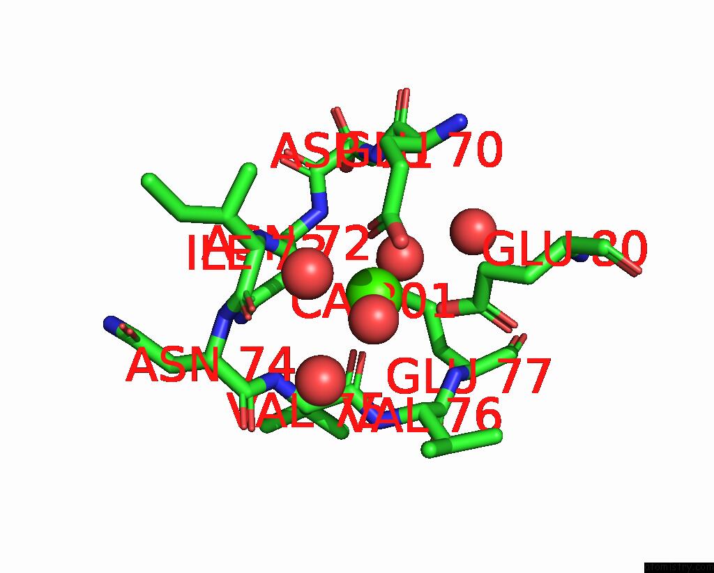
Mono view
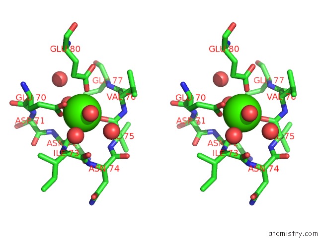
Stereo pair view
Mono view
Stereo pair view
A full contact list of Calcium with other atoms in the Ca binding
site number 1 of Crystal Structure of Beta-Trysin Complex in Cyclohexane within 5.0Å range:
|
Reference:
G.Zhu,
Q.Huang,
Y.Zhu,
Y.Li,
C.Chi,
Y.Tang.
X-Ray Study on An Artificial Mung Bean Inhibitor Complex with Bovine Beta-Trypsin in Neat Cyclohexane. Biochim.Biophys.Acta V.1546 98 2001.
ISSN: ISSN 0006-3002
PubMed: 11257512
DOI: 10.1016/S0167-4838(00)00299-5
Page generated: Mon Jul 7 15:11:40 2025
ISSN: ISSN 0006-3002
PubMed: 11257512
DOI: 10.1016/S0167-4838(00)00299-5
Last articles
Mg in 7BNRMg in 7BNK
Mg in 7BMC
Mg in 7BM9
Mg in 7BM8
Mg in 7BM6
Mg in 7BL4
Mg in 7BL6
Mg in 7BL5
Mg in 7BJT