Calcium »
PDB 1gk9-1gxo »
1gn1 »
Calcium in PDB 1gn1: Crystal Structure of the Mouse Cct Gamma Apical Domain (Monoclinic)
Protein crystallography data
The structure of Crystal Structure of the Mouse Cct Gamma Apical Domain (Monoclinic), PDB code: 1gn1
was solved by
G.Pappenberger,
J.A.Wilsher,
S.M.Roe,
K.R.Willison,
L.H.Pearl,
with X-Ray Crystallography technique. A brief refinement statistics is given in the table below:
| Resolution Low / High (Å) | 29.11 / 2.80 |
| Space group | P 1 21 1 |
| Cell size a, b, c (Å), α, β, γ (°) | 60.244, 234.229, 62.703, 90.00, 114.70, 90.00 |
| R / Rfree (%) | 23.8 / 28.5 |
Calcium Binding Sites:
The binding sites of Calcium atom in the Crystal Structure of the Mouse Cct Gamma Apical Domain (Monoclinic)
(pdb code 1gn1). This binding sites where shown within
5.0 Angstroms radius around Calcium atom.
In total 2 binding sites of Calcium where determined in the Crystal Structure of the Mouse Cct Gamma Apical Domain (Monoclinic), PDB code: 1gn1:
Jump to Calcium binding site number: 1; 2;
In total 2 binding sites of Calcium where determined in the Crystal Structure of the Mouse Cct Gamma Apical Domain (Monoclinic), PDB code: 1gn1:
Jump to Calcium binding site number: 1; 2;
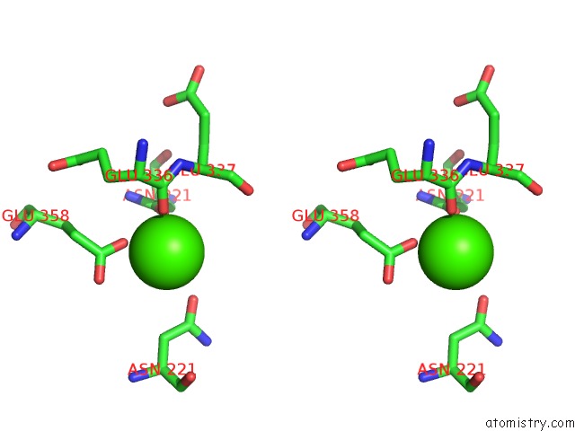
Calcium binding site 1 out of 2 in 1gn1
Go back to
Calcium binding site 1 out
of 2 in the Crystal Structure of the Mouse Cct Gamma Apical Domain (Monoclinic)

Mono view

Stereo pair view
Mono view
Stereo pair view
A full contact list of Calcium with other atoms in the Ca binding
site number 1 of Crystal Structure of the Mouse Cct Gamma Apical Domain (Monoclinic) within 5.0Å range:
|
Calcium binding site 2 out of 2 in 1gn1
Go back to
Calcium binding site 2 out
of 2 in the Crystal Structure of the Mouse Cct Gamma Apical Domain (Monoclinic)

Mono view
Stereo pair view
Mono view
Stereo pair view
A full contact list of Calcium with other atoms in the Ca binding
site number 2 of Crystal Structure of the Mouse Cct Gamma Apical Domain (Monoclinic) within 5.0Å range:
|
Reference:
G.Pappenberger,
J.A.Wilsher,
S.M.Roe,
D.J.Counsell,
K.R.Willison,
L.H.Pearl.
Crystal Structure of the Cct Gamma Apical Domain:: Implications For Substrate Binding to the Eukaryotic Cytosolic Chaperonin J.Mol.Biol. V. 318 1367 2002.
ISSN: ISSN 0022-2836
PubMed: 12083524
DOI: 10.1016/S0022-2836(02)00190-0
Page generated: Mon Jul 7 15:16:49 2025
ISSN: ISSN 0022-2836
PubMed: 12083524
DOI: 10.1016/S0022-2836(02)00190-0
Last articles
Mg in 4GMJMg in 4GNK
Mg in 4GNI
Mg in 4GN0
Mg in 4GMX
Mg in 4GME
Mg in 4GKM
Mg in 4GKR
Mg in 4GHL
Mg in 4GIU