Calcium »
PDB 1h80-1hny »
1hlu »
Calcium in PDB 1hlu: Structure of Bovine Beta-Actin-Profilin Complex with Actin Bound Atp Phosphates Solvent Accessible
Protein crystallography data
The structure of Structure of Bovine Beta-Actin-Profilin Complex with Actin Bound Atp Phosphates Solvent Accessible, PDB code: 1hlu
was solved by
J.K.Chik,
U.Lindberg,
C.E.Schutt,
with X-Ray Crystallography technique. A brief refinement statistics is given in the table below:
| Resolution Low / High (Å) | 8.00 / 2.65 |
| Space group | P 21 21 21 |
| Cell size a, b, c (Å), α, β, γ (°) | 38.140, 72.240, 185.700, 90.00, 90.00, 90.00 |
| R / Rfree (%) | 20.1 / 33 |
Calcium Binding Sites:
The binding sites of Calcium atom in the Structure of Bovine Beta-Actin-Profilin Complex with Actin Bound Atp Phosphates Solvent Accessible
(pdb code 1hlu). This binding sites where shown within
5.0 Angstroms radius around Calcium atom.
In total only one binding site of Calcium was determined in the Structure of Bovine Beta-Actin-Profilin Complex with Actin Bound Atp Phosphates Solvent Accessible, PDB code: 1hlu:
In total only one binding site of Calcium was determined in the Structure of Bovine Beta-Actin-Profilin Complex with Actin Bound Atp Phosphates Solvent Accessible, PDB code: 1hlu:
Calcium binding site 1 out of 1 in 1hlu
Go back to
Calcium binding site 1 out
of 1 in the Structure of Bovine Beta-Actin-Profilin Complex with Actin Bound Atp Phosphates Solvent Accessible
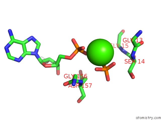
Mono view
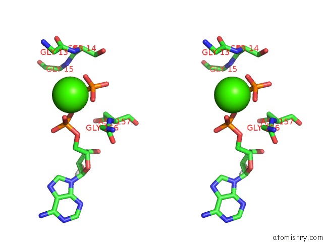
Stereo pair view
Mono view
Stereo pair view
A full contact list of Calcium with other atoms in the Ca binding
site number 1 of Structure of Bovine Beta-Actin-Profilin Complex with Actin Bound Atp Phosphates Solvent Accessible within 5.0Å range:
|
Reference:
J.K.Chik,
U.Lindberg,
C.E.Schutt.
The Structure of An Open State of Beta-Actin at 2.65 A Resolution. J.Mol.Biol. V. 263 607 1996.
ISSN: ISSN 0022-2836
PubMed: 8918942
DOI: 10.1006/JMBI.1996.0602
Page generated: Mon Jul 7 15:38:22 2025
ISSN: ISSN 0022-2836
PubMed: 8918942
DOI: 10.1006/JMBI.1996.0602
Last articles
Mg in 7VBZMg in 7VB6
Mg in 7VBN
Mg in 7VBW
Mg in 7VB7
Mg in 7VB5
Mg in 7VB3
Mg in 7VB4
Mg in 7V8I
Mg in 7V6K