Calcium »
PDB 1hov-1i6t »
1hv5 »
Calcium in PDB 1hv5: Crystal Structure of the Stromelysin-3 (Mmp-11) Catalytic Domain Complexed with A Phosphinic Inhibitor
Protein crystallography data
The structure of Crystal Structure of the Stromelysin-3 (Mmp-11) Catalytic Domain Complexed with A Phosphinic Inhibitor, PDB code: 1hv5
was solved by
A.L.Gall,
M.Ruff,
R.Kannan,
P.Cuniasse,
A.Yiotakis,
V.Dive,
M.C.Rio,
P.Basset,
D.Moras,
with X-Ray Crystallography technique. A brief refinement statistics is given in the table below:
| Resolution Low / High (Å) | 19.89 / 2.60 |
| Space group | P 21 21 21 |
| Cell size a, b, c (Å), α, β, γ (°) | 140.100, 148.500, 91.400, 90.00, 90.00, 90.00 |
| R / Rfree (%) | 21.8 / 26.2 |
Other elements in 1hv5:
The structure of Crystal Structure of the Stromelysin-3 (Mmp-11) Catalytic Domain Complexed with A Phosphinic Inhibitor also contains other interesting chemical elements:
| Zinc | (Zn) | 12 atoms |
Calcium Binding Sites:
The binding sites of Calcium atom in the Crystal Structure of the Stromelysin-3 (Mmp-11) Catalytic Domain Complexed with A Phosphinic Inhibitor
(pdb code 1hv5). This binding sites where shown within
5.0 Angstroms radius around Calcium atom.
In total 6 binding sites of Calcium where determined in the Crystal Structure of the Stromelysin-3 (Mmp-11) Catalytic Domain Complexed with A Phosphinic Inhibitor, PDB code: 1hv5:
Jump to Calcium binding site number: 1; 2; 3; 4; 5; 6;
In total 6 binding sites of Calcium where determined in the Crystal Structure of the Stromelysin-3 (Mmp-11) Catalytic Domain Complexed with A Phosphinic Inhibitor, PDB code: 1hv5:
Jump to Calcium binding site number: 1; 2; 3; 4; 5; 6;
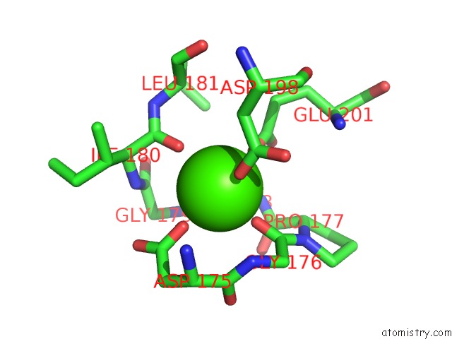
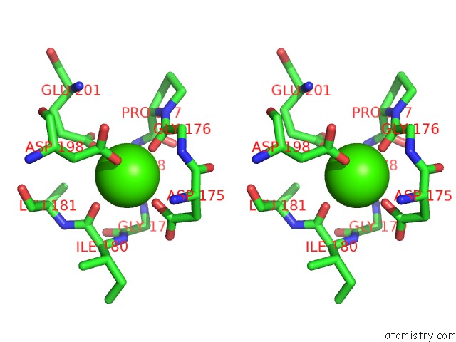
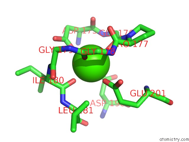
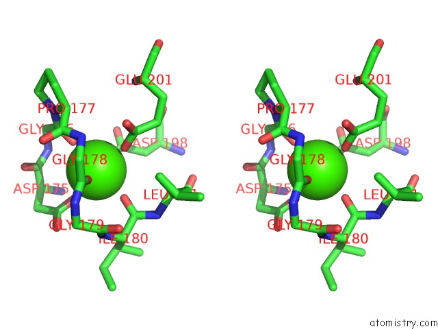
Calcium binding site 1 out of 6 in 1hv5
Go back to
Calcium binding site 1 out
of 6 in the Crystal Structure of the Stromelysin-3 (Mmp-11) Catalytic Domain Complexed with A Phosphinic Inhibitor
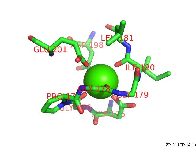
Mono view
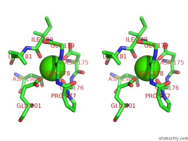
Stereo pair view
Mono view
Stereo pair view
A full contact list of Calcium with other atoms in the Ca binding
site number 1 of Crystal Structure of the Stromelysin-3 (Mmp-11) Catalytic Domain Complexed with A Phosphinic Inhibitor within 5.0Å range:
|
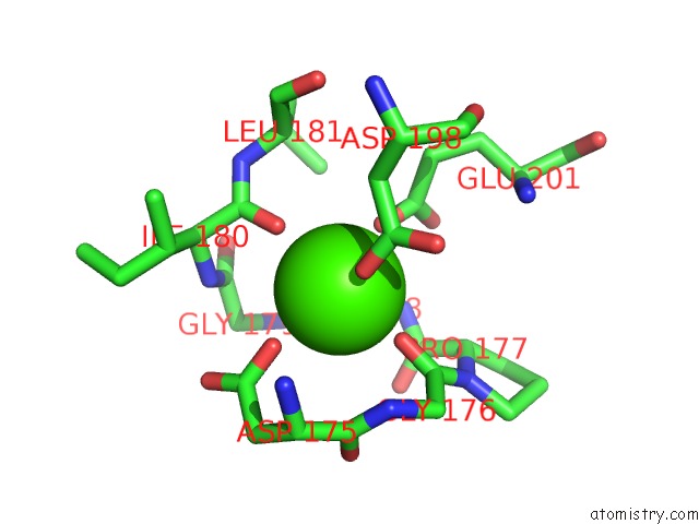
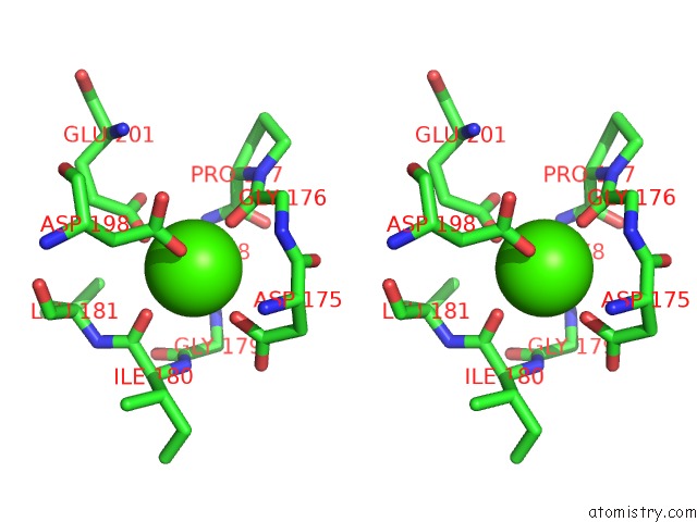
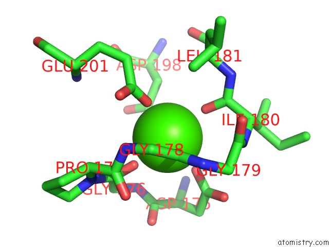
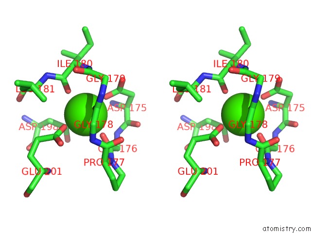
Calcium binding site 2 out of 6 in 1hv5
Go back to
Calcium binding site 2 out
of 6 in the Crystal Structure of the Stromelysin-3 (Mmp-11) Catalytic Domain Complexed with A Phosphinic Inhibitor
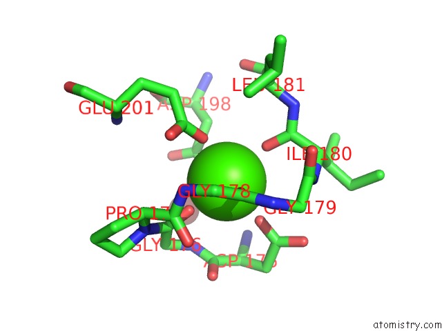
Mono view
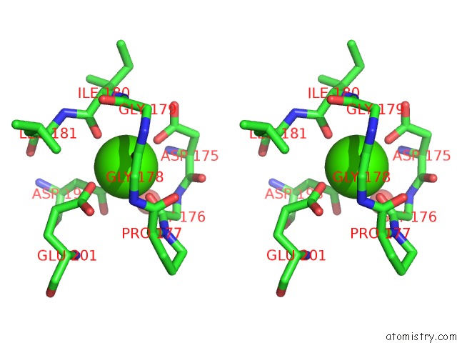
Stereo pair view
Mono view
Stereo pair view
A full contact list of Calcium with other atoms in the Ca binding
site number 2 of Crystal Structure of the Stromelysin-3 (Mmp-11) Catalytic Domain Complexed with A Phosphinic Inhibitor within 5.0Å range:
|
Calcium binding site 3 out of 6 in 1hv5
Go back to
Calcium binding site 3 out
of 6 in the Crystal Structure of the Stromelysin-3 (Mmp-11) Catalytic Domain Complexed with A Phosphinic Inhibitor
Mono view
Stereo pair view
Mono view
Stereo pair view
A full contact list of Calcium with other atoms in the Ca binding
site number 3 of Crystal Structure of the Stromelysin-3 (Mmp-11) Catalytic Domain Complexed with A Phosphinic Inhibitor within 5.0Å range:
|
Calcium binding site 4 out of 6 in 1hv5
Go back to
Calcium binding site 4 out
of 6 in the Crystal Structure of the Stromelysin-3 (Mmp-11) Catalytic Domain Complexed with A Phosphinic Inhibitor
Mono view
Stereo pair view
Mono view
Stereo pair view
A full contact list of Calcium with other atoms in the Ca binding
site number 4 of Crystal Structure of the Stromelysin-3 (Mmp-11) Catalytic Domain Complexed with A Phosphinic Inhibitor within 5.0Å range:
|
Calcium binding site 5 out of 6 in 1hv5
Go back to
Calcium binding site 5 out
of 6 in the Crystal Structure of the Stromelysin-3 (Mmp-11) Catalytic Domain Complexed with A Phosphinic Inhibitor
Mono view
Stereo pair view
Mono view
Stereo pair view
A full contact list of Calcium with other atoms in the Ca binding
site number 5 of Crystal Structure of the Stromelysin-3 (Mmp-11) Catalytic Domain Complexed with A Phosphinic Inhibitor within 5.0Å range:
|
Calcium binding site 6 out of 6 in 1hv5
Go back to
Calcium binding site 6 out
of 6 in the Crystal Structure of the Stromelysin-3 (Mmp-11) Catalytic Domain Complexed with A Phosphinic Inhibitor
Mono view
Stereo pair view
Mono view
Stereo pair view
A full contact list of Calcium with other atoms in the Ca binding
site number 6 of Crystal Structure of the Stromelysin-3 (Mmp-11) Catalytic Domain Complexed with A Phosphinic Inhibitor within 5.0Å range:
|
Reference:
A.L.Gall,
M.Ruff,
R.Kannan,
P.Cuniasse,
A.Yiotakis,
V.Dive,
M.C.Rio,
P.Basset,
D.Moras.
Crystal Structure of the Stromelysin-3 (Mmp-11) Catalytic Domain Complexed with A Phosphinic Inhibitor Mimicking the Transition-State. J.Mol.Biol. V. 307 577 2001.
ISSN: ISSN 0022-2836
PubMed: 11254383
DOI: 10.1006/JMBI.2001.4493
Page generated: Mon Jul 7 15:42:26 2025
ISSN: ISSN 0022-2836
PubMed: 11254383
DOI: 10.1006/JMBI.2001.4493
Last articles
Mg in 4NO6Mg in 4NPV
Mg in 4NOB
Mg in 4NNW
Mg in 4NO1
Mg in 4NNN
Mg in 4NO4
Mg in 4NNB
Mg in 4NMN
Mg in 4NNA