Calcium »
PDB 1i73-1iqi »
1ic6 »
Calcium in PDB 1ic6: Structure of A Serine Protease Proteinase K From Tritirachium Album Limber at 0.98 A Resolution
Enzymatic activity of Structure of A Serine Protease Proteinase K From Tritirachium Album Limber at 0.98 A Resolution
All present enzymatic activity of Structure of A Serine Protease Proteinase K From Tritirachium Album Limber at 0.98 A Resolution:
3.4.21.64;
3.4.21.64;
Protein crystallography data
The structure of Structure of A Serine Protease Proteinase K From Tritirachium Album Limber at 0.98 A Resolution, PDB code: 1ic6
was solved by
C.Betzel,
S.Gourinath,
P.Kumar,
P.Kaur,
M.Perbandt,
S.Eschenburg,
T.P.Singh,
with X-Ray Crystallography technique. A brief refinement statistics is given in the table below:
| Resolution Low / High (Å) | 10.00 / 0.98 |
| Space group | P 43 21 2 |
| Cell size a, b, c (Å), α, β, γ (°) | 67.288, 67.288, 106.582, 90.00, 90.00, 90.00 |
| R / Rfree (%) | 11.4 / 12.4 |
Calcium Binding Sites:
The binding sites of Calcium atom in the Structure of A Serine Protease Proteinase K From Tritirachium Album Limber at 0.98 A Resolution
(pdb code 1ic6). This binding sites where shown within
5.0 Angstroms radius around Calcium atom.
In total 2 binding sites of Calcium where determined in the Structure of A Serine Protease Proteinase K From Tritirachium Album Limber at 0.98 A Resolution, PDB code: 1ic6:
Jump to Calcium binding site number: 1; 2;
In total 2 binding sites of Calcium where determined in the Structure of A Serine Protease Proteinase K From Tritirachium Album Limber at 0.98 A Resolution, PDB code: 1ic6:
Jump to Calcium binding site number: 1; 2;
Calcium binding site 1 out of 2 in 1ic6
Go back to
Calcium binding site 1 out
of 2 in the Structure of A Serine Protease Proteinase K From Tritirachium Album Limber at 0.98 A Resolution
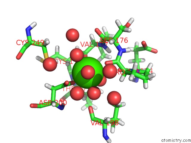
Mono view
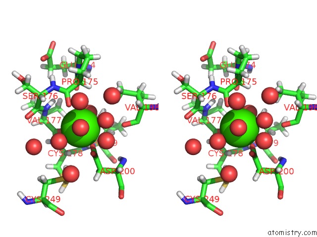
Stereo pair view
Mono view
Stereo pair view
A full contact list of Calcium with other atoms in the Ca binding
site number 1 of Structure of A Serine Protease Proteinase K From Tritirachium Album Limber at 0.98 A Resolution within 5.0Å range:
|
Calcium binding site 2 out of 2 in 1ic6
Go back to
Calcium binding site 2 out
of 2 in the Structure of A Serine Protease Proteinase K From Tritirachium Album Limber at 0.98 A Resolution
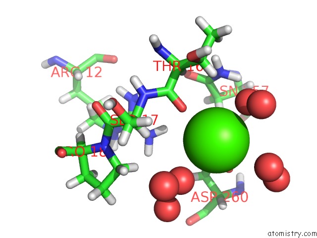
Mono view
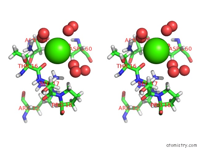
Stereo pair view
Mono view
Stereo pair view
A full contact list of Calcium with other atoms in the Ca binding
site number 2 of Structure of A Serine Protease Proteinase K From Tritirachium Album Limber at 0.98 A Resolution within 5.0Å range:
|
Reference:
C.Betzel,
S.Gourinath,
P.Kumar,
P.Kaur,
M.Perbandt,
S.Eschenburg,
T.P.Singh.
Structure of A Serine Protease Proteinase K From Tritirachium Album Limber at 0.98 A Resolution. Biochemistry V. 40 3080 2001.
ISSN: ISSN 0006-2960
PubMed: 11258922
DOI: 10.1021/BI002538N
Page generated: Mon Jul 7 15:49:09 2025
ISSN: ISSN 0006-2960
PubMed: 11258922
DOI: 10.1021/BI002538N
Last articles
Mg in 6CA4Mg in 6C90
Mg in 6CA0
Mg in 6C9Y
Mg in 6C8Z
Mg in 6C8P
Mg in 6C8N
Mg in 6C8O
Mg in 6C8D
Mg in 6C8L