Calcium »
PDB 1i73-1iqi »
1ika »
Calcium in PDB 1ika: Structure of Isocitrate Dehydrogenase with Alpha-Ketoglutarate at 2.7 Angstroms Resolution: Conformational Changes Induced By Decarboxylation of Isocitrate
Enzymatic activity of Structure of Isocitrate Dehydrogenase with Alpha-Ketoglutarate at 2.7 Angstroms Resolution: Conformational Changes Induced By Decarboxylation of Isocitrate
All present enzymatic activity of Structure of Isocitrate Dehydrogenase with Alpha-Ketoglutarate at 2.7 Angstroms Resolution: Conformational Changes Induced By Decarboxylation of Isocitrate:
1.1.1.42;
1.1.1.42;
Protein crystallography data
The structure of Structure of Isocitrate Dehydrogenase with Alpha-Ketoglutarate at 2.7 Angstroms Resolution: Conformational Changes Induced By Decarboxylation of Isocitrate, PDB code: 1ika
was solved by
B.L.Stoddard,
D.E.Koshland Junior,
with X-Ray Crystallography technique. A brief refinement statistics is given in the table below:
| Resolution Low / High (Å) | 30.00 / 2.70 |
| Space group | P 43 21 2 |
| Cell size a, b, c (Å), α, β, γ (°) | 105.100, 105.100, 150.300, 90.00, 90.00, 90.00 |
| R / Rfree (%) | n/a / n/a |
Calcium Binding Sites:
The binding sites of Calcium atom in the Structure of Isocitrate Dehydrogenase with Alpha-Ketoglutarate at 2.7 Angstroms Resolution: Conformational Changes Induced By Decarboxylation of Isocitrate
(pdb code 1ika). This binding sites where shown within
5.0 Angstroms radius around Calcium atom.
In total only one binding site of Calcium was determined in the Structure of Isocitrate Dehydrogenase with Alpha-Ketoglutarate at 2.7 Angstroms Resolution: Conformational Changes Induced By Decarboxylation of Isocitrate, PDB code: 1ika:
In total only one binding site of Calcium was determined in the Structure of Isocitrate Dehydrogenase with Alpha-Ketoglutarate at 2.7 Angstroms Resolution: Conformational Changes Induced By Decarboxylation of Isocitrate, PDB code: 1ika:
Calcium binding site 1 out of 1 in 1ika
Go back to
Calcium binding site 1 out
of 1 in the Structure of Isocitrate Dehydrogenase with Alpha-Ketoglutarate at 2.7 Angstroms Resolution: Conformational Changes Induced By Decarboxylation of Isocitrate
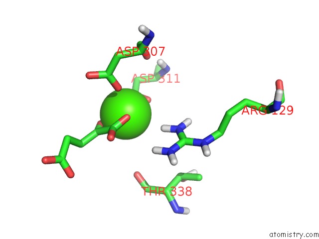
Mono view
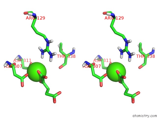
Stereo pair view
Mono view
Stereo pair view
A full contact list of Calcium with other atoms in the Ca binding
site number 1 of Structure of Isocitrate Dehydrogenase with Alpha-Ketoglutarate at 2.7 Angstroms Resolution: Conformational Changes Induced By Decarboxylation of Isocitrate within 5.0Å range:
|
Reference:
B.L.Stoddard,
D.E.Koshland Jr..
Structure of Isocitrate Dehydrogenase with Alpha-Ketoglutarate at 2.7-A Resolution: Conformational Changes Induced By Decarboxylation of Isocitrate. Biochemistry V. 32 9317 1993.
ISSN: ISSN 0006-2960
PubMed: 8369301
DOI: 10.1021/BI00087A009
Page generated: Mon Jul 7 15:50:28 2025
ISSN: ISSN 0006-2960
PubMed: 8369301
DOI: 10.1021/BI00087A009
Last articles
Mg in 6CA4Mg in 6C90
Mg in 6CA0
Mg in 6C9Y
Mg in 6C8Z
Mg in 6C8P
Mg in 6C8N
Mg in 6C8O
Mg in 6C8D
Mg in 6C8L