Calcium »
PDB 1jf5-1jv2 »
1jhn »
Calcium in PDB 1jhn: Crystal Structure of the Lumenal Domain of Calnexin
Protein crystallography data
The structure of Crystal Structure of the Lumenal Domain of Calnexin, PDB code: 1jhn
was solved by
J.D.Schrag,
J.M.Bergeron,
Y.Li,
S.Borisova,
M.Hahn,
D.Y.Thomas,
M.Cygler,
with X-Ray Crystallography technique. A brief refinement statistics is given in the table below:
| Resolution Low / High (Å) | 33.80 / 2.90 |
| Space group | P 43 21 2 |
| Cell size a, b, c (Å), α, β, γ (°) | 85.772, 85.772, 143.476, 90.00, 90.00, 90.00 |
| R / Rfree (%) | 32.7 / 37.7 |
Calcium Binding Sites:
The binding sites of Calcium atom in the Crystal Structure of the Lumenal Domain of Calnexin
(pdb code 1jhn). This binding sites where shown within
5.0 Angstroms radius around Calcium atom.
In total only one binding site of Calcium was determined in the Crystal Structure of the Lumenal Domain of Calnexin, PDB code: 1jhn:
In total only one binding site of Calcium was determined in the Crystal Structure of the Lumenal Domain of Calnexin, PDB code: 1jhn:
Calcium binding site 1 out of 1 in 1jhn
Go back to
Calcium binding site 1 out
of 1 in the Crystal Structure of the Lumenal Domain of Calnexin
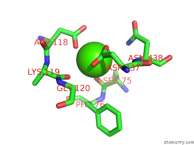
Mono view
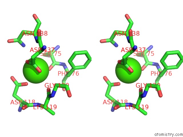
Stereo pair view
Mono view
Stereo pair view
A full contact list of Calcium with other atoms in the Ca binding
site number 1 of Crystal Structure of the Lumenal Domain of Calnexin within 5.0Å range:
|
Reference:
J.D.Schrag,
J.J.Bergeron,
Y.Li,
S.Borisova,
M.Hahn,
D.Y.Thomas,
M.Cygler.
The Structure of Calnexin, An Er Chaperone Involved in Quality Control of Protein Folding. Mol.Cell V. 8 633 2001.
ISSN: ISSN 1097-2765
PubMed: 11583625
DOI: 10.1016/S1097-2765(01)00318-5
Page generated: Mon Jul 7 16:08:09 2025
ISSN: ISSN 1097-2765
PubMed: 11583625
DOI: 10.1016/S1097-2765(01)00318-5
Last articles
Mg in 7ADEMg in 7ADI
Mg in 7ADD
Mg in 7ADC
Mg in 7ADB
Mg in 7AD9
Mg in 7ACQ
Mg in 7ACF
Mg in 7ACA
Mg in 7ACH