Calcium »
PDB 1k9u-1kre »
1kek »
Calcium in PDB 1kek: Crystal Structure of the Free Radical Intermediate of Pyruvate:Ferredoxin Oxidoreductase
Enzymatic activity of Crystal Structure of the Free Radical Intermediate of Pyruvate:Ferredoxin Oxidoreductase
All present enzymatic activity of Crystal Structure of the Free Radical Intermediate of Pyruvate:Ferredoxin Oxidoreductase:
1.2.7.1;
1.2.7.1;
Protein crystallography data
The structure of Crystal Structure of the Free Radical Intermediate of Pyruvate:Ferredoxin Oxidoreductase, PDB code: 1kek
was solved by
E.Chabriere,
X.Vernede,
B.Guigliarelli,
M.-H.Charon,
E.C.Hatchikian,
J.C.Fontecilla-Camps,
with X-Ray Crystallography technique. A brief refinement statistics is given in the table below:
| Resolution Low / High (Å) | 27.38 / 1.90 |
| Space group | P 21 21 21 |
| Cell size a, b, c (Å), α, β, γ (°) | 86.110, 145.760, 210.260, 90.00, 90.00, 90.00 |
| R / Rfree (%) | 17.8 / 22.7 |
Other elements in 1kek:
The structure of Crystal Structure of the Free Radical Intermediate of Pyruvate:Ferredoxin Oxidoreductase also contains other interesting chemical elements:
| Magnesium | (Mg) | 2 atoms |
| Iron | (Fe) | 24 atoms |
Calcium Binding Sites:
The binding sites of Calcium atom in the Crystal Structure of the Free Radical Intermediate of Pyruvate:Ferredoxin Oxidoreductase
(pdb code 1kek). This binding sites where shown within
5.0 Angstroms radius around Calcium atom.
In total 2 binding sites of Calcium where determined in the Crystal Structure of the Free Radical Intermediate of Pyruvate:Ferredoxin Oxidoreductase, PDB code: 1kek:
Jump to Calcium binding site number: 1; 2;
In total 2 binding sites of Calcium where determined in the Crystal Structure of the Free Radical Intermediate of Pyruvate:Ferredoxin Oxidoreductase, PDB code: 1kek:
Jump to Calcium binding site number: 1; 2;
Calcium binding site 1 out of 2 in 1kek
Go back to
Calcium binding site 1 out
of 2 in the Crystal Structure of the Free Radical Intermediate of Pyruvate:Ferredoxin Oxidoreductase
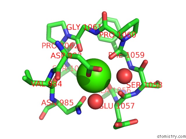
Mono view
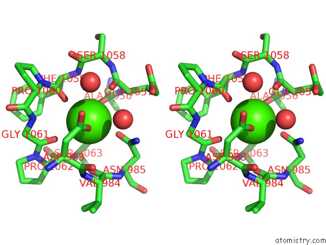
Stereo pair view
Mono view
Stereo pair view
A full contact list of Calcium with other atoms in the Ca binding
site number 1 of Crystal Structure of the Free Radical Intermediate of Pyruvate:Ferredoxin Oxidoreductase within 5.0Å range:
|
Calcium binding site 2 out of 2 in 1kek
Go back to
Calcium binding site 2 out
of 2 in the Crystal Structure of the Free Radical Intermediate of Pyruvate:Ferredoxin Oxidoreductase
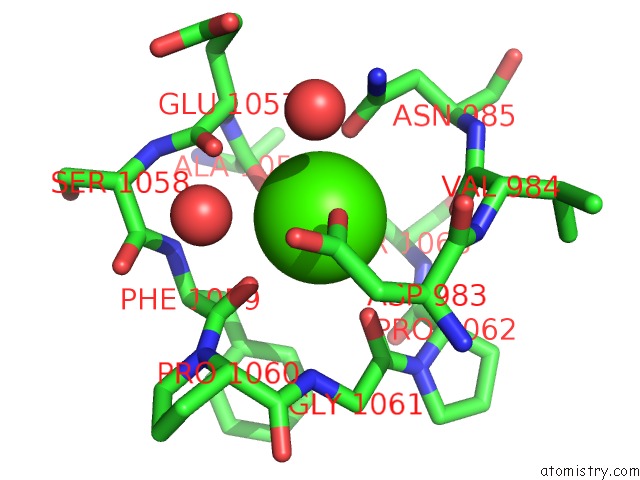
Mono view
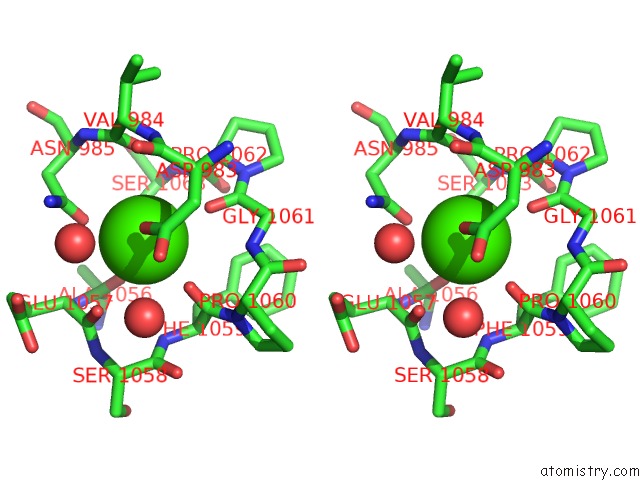
Stereo pair view
Mono view
Stereo pair view
A full contact list of Calcium with other atoms in the Ca binding
site number 2 of Crystal Structure of the Free Radical Intermediate of Pyruvate:Ferredoxin Oxidoreductase within 5.0Å range:
|
Reference:
E.Chabriere,
X.Vernede,
B.Guigliarelli,
M.H.Charon,
E.C.Hatchikian,
J.C.Fontecilla-Camps.
Crystal Structure of the Free Radical Intermediate of Pyruvate:Ferredoxin Oxidoreductase. Science V. 294 2559 2001.
ISSN: ISSN 0036-8075
PubMed: 11752578
DOI: 10.1126/SCIENCE.1066198
Page generated: Mon Jul 7 16:28:13 2025
ISSN: ISSN 0036-8075
PubMed: 11752578
DOI: 10.1126/SCIENCE.1066198
Last articles
Fe in 8VOTFe in 8VOK
Fe in 8VOC
Fe in 8RQZ
Fe in 8RTM
Fe in 8RTL
F in 9RF5
F in 9N6R
F in 9N6P
F in 9N6M