Calcium »
PDB 1llp-1ltj »
1low »
Calcium in PDB 1low: X-Ray Structure of the H40A Mutant of Ribonuclease T1 Complexed with 3'-Guanosine Monophosphate
Enzymatic activity of X-Ray Structure of the H40A Mutant of Ribonuclease T1 Complexed with 3'-Guanosine Monophosphate
All present enzymatic activity of X-Ray Structure of the H40A Mutant of Ribonuclease T1 Complexed with 3'-Guanosine Monophosphate:
3.1.27.3;
3.1.27.3;
Protein crystallography data
The structure of X-Ray Structure of the H40A Mutant of Ribonuclease T1 Complexed with 3'-Guanosine Monophosphate, PDB code: 1low
was solved by
P.Mignon,
J.Steyaert,
R.Loris,
P.Geerlings,
S.Loverix,
with X-Ray Crystallography technique. A brief refinement statistics is given in the table below:
| Resolution Low / High (Å) | 22.80 / 1.90 |
| Space group | P 21 21 21 |
| Cell size a, b, c (Å), α, β, γ (°) | 40.217, 45.685, 50.136, 90.00, 90.00, 90.00 |
| R / Rfree (%) | 20.6 / 22.9 |
Calcium Binding Sites:
The binding sites of Calcium atom in the X-Ray Structure of the H40A Mutant of Ribonuclease T1 Complexed with 3'-Guanosine Monophosphate
(pdb code 1low). This binding sites where shown within
5.0 Angstroms radius around Calcium atom.
In total only one binding site of Calcium was determined in the X-Ray Structure of the H40A Mutant of Ribonuclease T1 Complexed with 3'-Guanosine Monophosphate, PDB code: 1low:
In total only one binding site of Calcium was determined in the X-Ray Structure of the H40A Mutant of Ribonuclease T1 Complexed with 3'-Guanosine Monophosphate, PDB code: 1low:
Calcium binding site 1 out of 1 in 1low
Go back to
Calcium binding site 1 out
of 1 in the X-Ray Structure of the H40A Mutant of Ribonuclease T1 Complexed with 3'-Guanosine Monophosphate
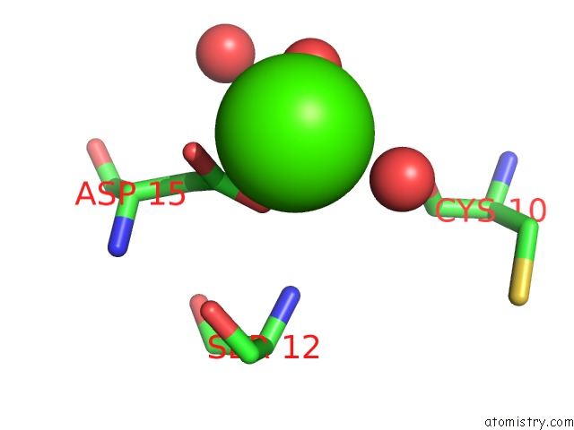
Mono view
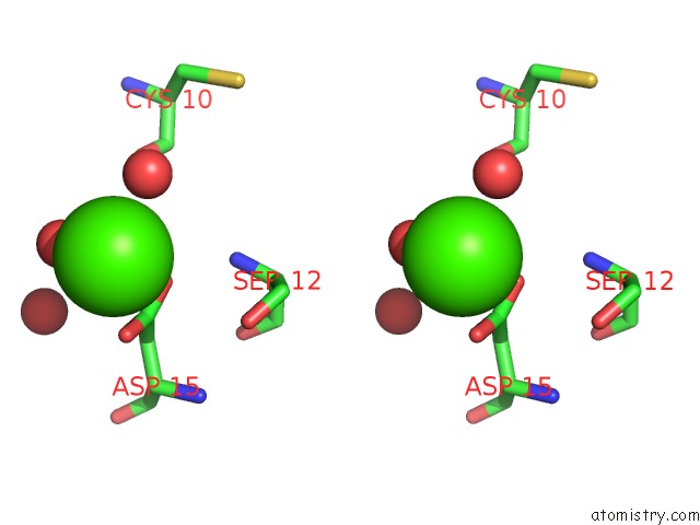
Stereo pair view
Mono view
Stereo pair view
A full contact list of Calcium with other atoms in the Ca binding
site number 1 of X-Ray Structure of the H40A Mutant of Ribonuclease T1 Complexed with 3'-Guanosine Monophosphate within 5.0Å range:
|
Reference:
P.Mignon,
J.Steyaert,
R.Loris,
P.Geerlings,
S.Loverix.
A Nucleophile Activation Dyad in Ribonucleases. A Combined X-Ray Crystallographic/Ab Initio Quantum Chemical Study J.Biol.Chem. V. 277 36770 2002.
ISSN: ISSN 0021-9258
PubMed: 12122018
DOI: 10.1074/JBC.M206461200
Page generated: Mon Jul 7 16:57:41 2025
ISSN: ISSN 0021-9258
PubMed: 12122018
DOI: 10.1074/JBC.M206461200
Last articles
K in 6ASOK in 6AFZ
K in 6AFY
K in 6AFX
K in 6AFW
K in 6AFV
K in 6AFU
K in 6AFT
K in 6AFS
K in 6ABM