Calcium »
PDB 1llp-1ltj »
1ltj »
Calcium in PDB 1ltj: Crystal Structure of Recombinant Human Fibrinogen Fragment D with the Peptide Ligands Gly-Pro-Arg-Pro-Amide and Gly-His-Arg-Pro-Amide
Protein crystallography data
The structure of Crystal Structure of Recombinant Human Fibrinogen Fragment D with the Peptide Ligands Gly-Pro-Arg-Pro-Amide and Gly-His-Arg-Pro-Amide, PDB code: 1ltj
was solved by
M.S.Kostelansky,
L.Betts,
O.V.Gorkun,
S.T.Lord,
with X-Ray Crystallography technique. A brief refinement statistics is given in the table below:
| Resolution Low / High (Å) | 18.02 / 2.80 |
| Space group | P 21 21 21 |
| Cell size a, b, c (Å), α, β, γ (°) | 89.284, 94.218, 226.936, 90.00, 90.00, 90.00 |
| R / Rfree (%) | 21.2 / 27 |
Calcium Binding Sites:
The binding sites of Calcium atom in the Crystal Structure of Recombinant Human Fibrinogen Fragment D with the Peptide Ligands Gly-Pro-Arg-Pro-Amide and Gly-His-Arg-Pro-Amide
(pdb code 1ltj). This binding sites where shown within
5.0 Angstroms radius around Calcium atom.
In total 4 binding sites of Calcium where determined in the Crystal Structure of Recombinant Human Fibrinogen Fragment D with the Peptide Ligands Gly-Pro-Arg-Pro-Amide and Gly-His-Arg-Pro-Amide, PDB code: 1ltj:
Jump to Calcium binding site number: 1; 2; 3; 4;
In total 4 binding sites of Calcium where determined in the Crystal Structure of Recombinant Human Fibrinogen Fragment D with the Peptide Ligands Gly-Pro-Arg-Pro-Amide and Gly-His-Arg-Pro-Amide, PDB code: 1ltj:
Jump to Calcium binding site number: 1; 2; 3; 4;
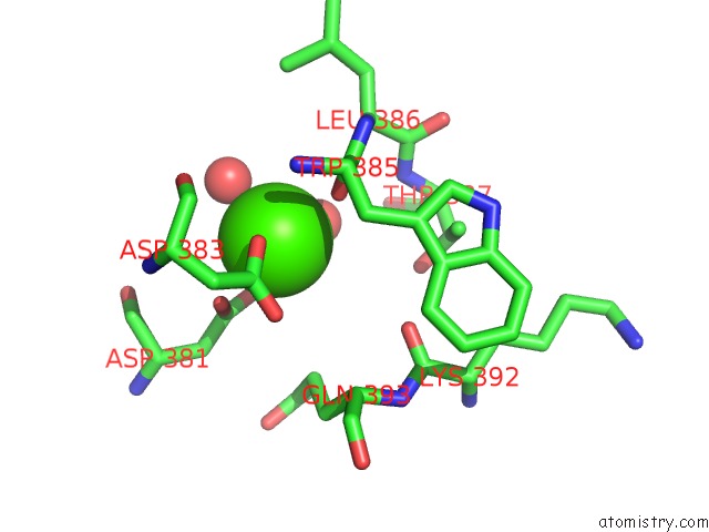
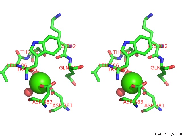
Calcium binding site 1 out of 4 in 1ltj
Go back to
Calcium binding site 1 out
of 4 in the Crystal Structure of Recombinant Human Fibrinogen Fragment D with the Peptide Ligands Gly-Pro-Arg-Pro-Amide and Gly-His-Arg-Pro-Amide
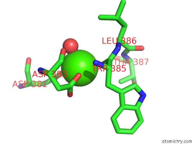
Mono view
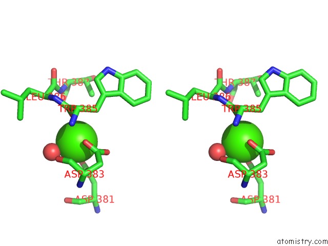
Stereo pair view
Mono view
Stereo pair view
A full contact list of Calcium with other atoms in the Ca binding
site number 1 of Crystal Structure of Recombinant Human Fibrinogen Fragment D with the Peptide Ligands Gly-Pro-Arg-Pro-Amide and Gly-His-Arg-Pro-Amide within 5.0Å range:
|
Calcium binding site 2 out of 4 in 1ltj
Go back to
Calcium binding site 2 out
of 4 in the Crystal Structure of Recombinant Human Fibrinogen Fragment D with the Peptide Ligands Gly-Pro-Arg-Pro-Amide and Gly-His-Arg-Pro-Amide
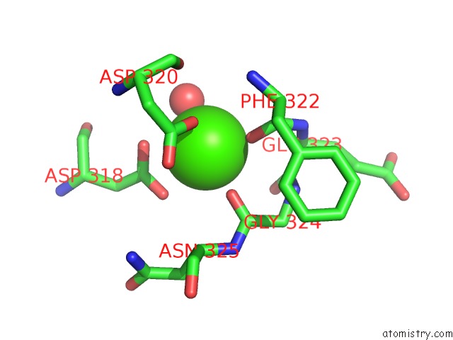
Mono view
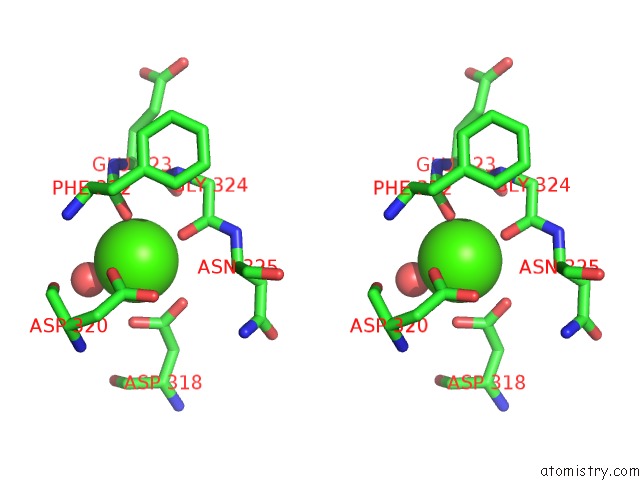
Stereo pair view
Mono view
Stereo pair view
A full contact list of Calcium with other atoms in the Ca binding
site number 2 of Crystal Structure of Recombinant Human Fibrinogen Fragment D with the Peptide Ligands Gly-Pro-Arg-Pro-Amide and Gly-His-Arg-Pro-Amide within 5.0Å range:
|
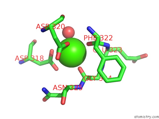
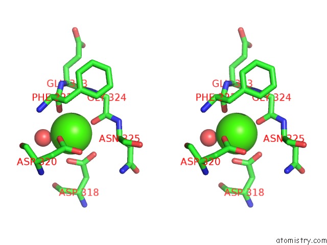
Calcium binding site 3 out of 4 in 1ltj
Go back to
Calcium binding site 3 out
of 4 in the Crystal Structure of Recombinant Human Fibrinogen Fragment D with the Peptide Ligands Gly-Pro-Arg-Pro-Amide and Gly-His-Arg-Pro-Amide
Mono view
Stereo pair view
Mono view
Stereo pair view
A full contact list of Calcium with other atoms in the Ca binding
site number 3 of Crystal Structure of Recombinant Human Fibrinogen Fragment D with the Peptide Ligands Gly-Pro-Arg-Pro-Amide and Gly-His-Arg-Pro-Amide within 5.0Å range:
|
Calcium binding site 4 out of 4 in 1ltj
Go back to
Calcium binding site 4 out
of 4 in the Crystal Structure of Recombinant Human Fibrinogen Fragment D with the Peptide Ligands Gly-Pro-Arg-Pro-Amide and Gly-His-Arg-Pro-Amide
Mono view
Stereo pair view
Mono view
Stereo pair view
A full contact list of Calcium with other atoms in the Ca binding
site number 4 of Crystal Structure of Recombinant Human Fibrinogen Fragment D with the Peptide Ligands Gly-Pro-Arg-Pro-Amide and Gly-His-Arg-Pro-Amide within 5.0Å range:
|
Reference:
M.S.Kostelansky,
L.Betts,
O.V.Gorkun,
S.T.Lord.
2.8 A Crystal Structures of Recombinant Fibrinogen Fragment D with and Without Two Peptide Ligands: Ghrp Binding to the "B" Site Disrupts Its Nearby Calcium-Binding Site. Biochemistry V. 41 12124 2002.
ISSN: ISSN 0006-2960
PubMed: 12356313
DOI: 10.1021/BI0261894
Page generated: Mon Jul 7 16:59:11 2025
ISSN: ISSN 0006-2960
PubMed: 12356313
DOI: 10.1021/BI0261894
Last articles
Mg in 3G73Mg in 3G37
Mg in 3G6Y
Mg in 3G6X
Mg in 3G6W
Mg in 3G6V
Mg in 3G6K
Mg in 3G5A
Mg in 3G5S
Mg in 3G58