Calcium »
PDB 1lu1-1m8v »
1lv8 »
Calcium in PDB 1lv8: Crystal Structure of Calf Spleen Purine Nucleoside Phosphorylase in A New Space Group with Full Trimer in the Asymmetric Unit
Enzymatic activity of Crystal Structure of Calf Spleen Purine Nucleoside Phosphorylase in A New Space Group with Full Trimer in the Asymmetric Unit
All present enzymatic activity of Crystal Structure of Calf Spleen Purine Nucleoside Phosphorylase in A New Space Group with Full Trimer in the Asymmetric Unit:
2.4.2.1;
2.4.2.1;
Protein crystallography data
The structure of Crystal Structure of Calf Spleen Purine Nucleoside Phosphorylase in A New Space Group with Full Trimer in the Asymmetric Unit, PDB code: 1lv8
was solved by
A.Bzowska,
G.Koellner,
B.Wielgus-Kutrowska,
A.Stroh,
G.Raszewski,
A.Holy,
T.Steiner,
J.Frank,
with X-Ray Crystallography technique. A brief refinement statistics is given in the table below:
| Resolution Low / High (Å) | 19.85 / 2.30 |
| Space group | P 21 21 21 |
| Cell size a, b, c (Å), α, β, γ (°) | 79.140, 134.235, 177.148, 90.00, 90.00, 90.00 |
| R / Rfree (%) | 18.9 / 25.8 |
Calcium Binding Sites:
The binding sites of Calcium atom in the Crystal Structure of Calf Spleen Purine Nucleoside Phosphorylase in A New Space Group with Full Trimer in the Asymmetric Unit
(pdb code 1lv8). This binding sites where shown within
5.0 Angstroms radius around Calcium atom.
In total 6 binding sites of Calcium where determined in the Crystal Structure of Calf Spleen Purine Nucleoside Phosphorylase in A New Space Group with Full Trimer in the Asymmetric Unit, PDB code: 1lv8:
Jump to Calcium binding site number: 1; 2; 3; 4; 5; 6;
In total 6 binding sites of Calcium where determined in the Crystal Structure of Calf Spleen Purine Nucleoside Phosphorylase in A New Space Group with Full Trimer in the Asymmetric Unit, PDB code: 1lv8:
Jump to Calcium binding site number: 1; 2; 3; 4; 5; 6;
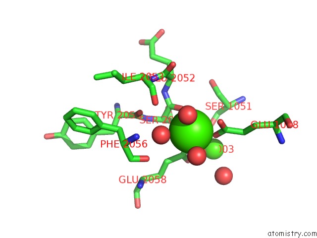
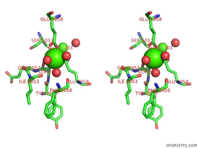
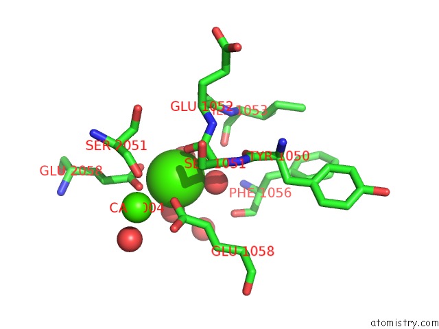
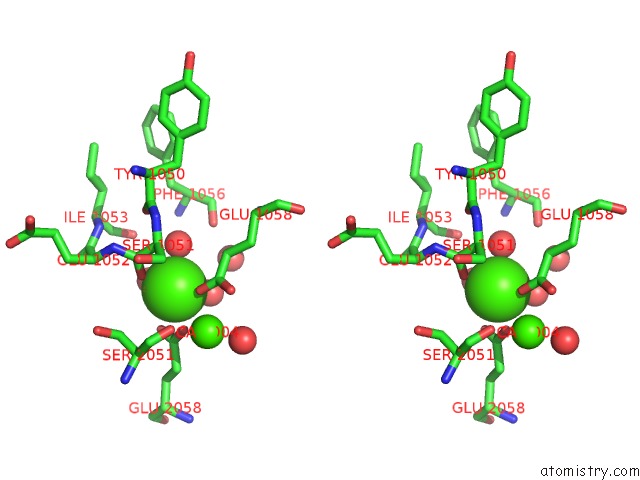
Calcium binding site 1 out of 6 in 1lv8
Go back to
Calcium binding site 1 out
of 6 in the Crystal Structure of Calf Spleen Purine Nucleoside Phosphorylase in A New Space Group with Full Trimer in the Asymmetric Unit
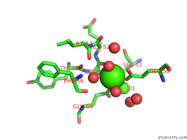
Mono view
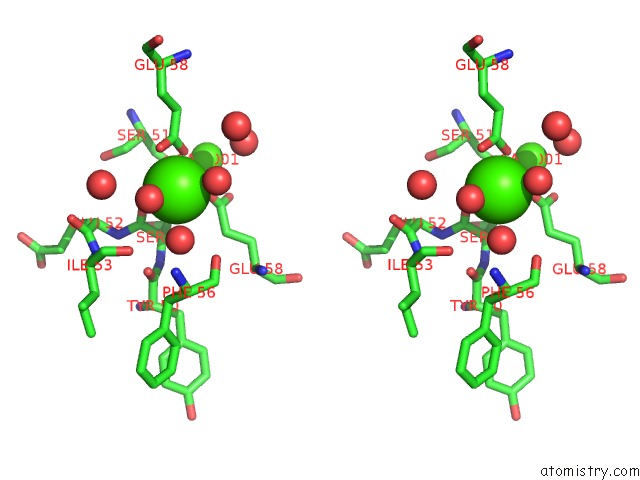
Stereo pair view
Mono view
Stereo pair view
A full contact list of Calcium with other atoms in the Ca binding
site number 1 of Crystal Structure of Calf Spleen Purine Nucleoside Phosphorylase in A New Space Group with Full Trimer in the Asymmetric Unit within 5.0Å range:
|
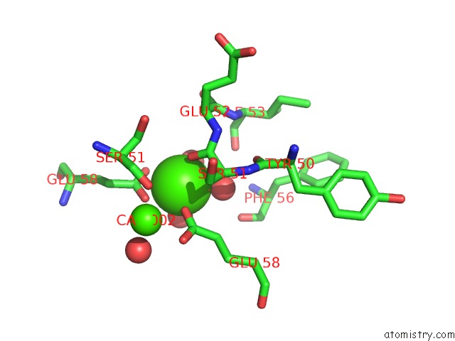
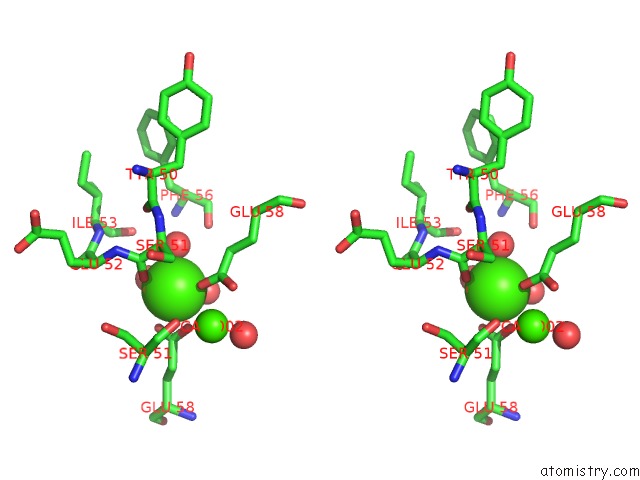
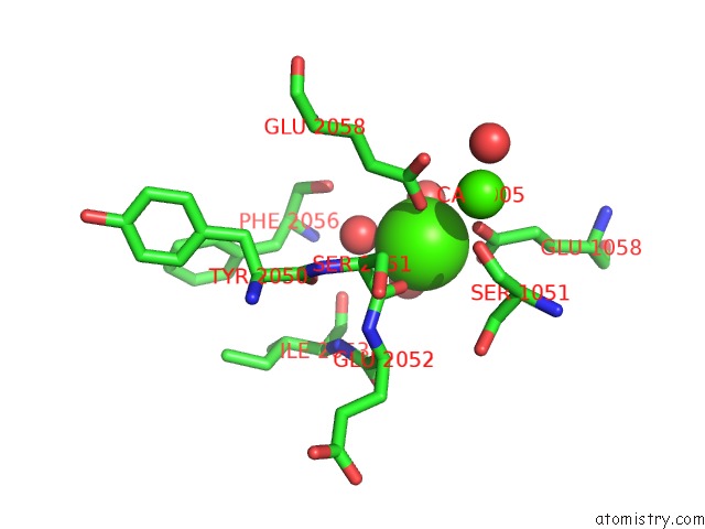
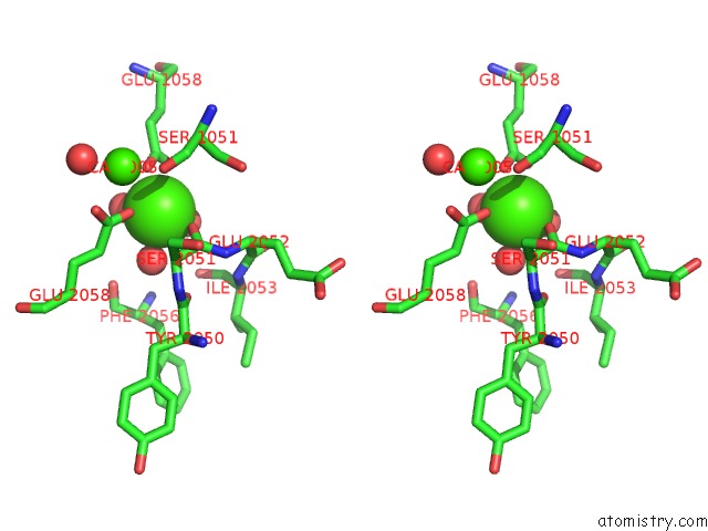
Calcium binding site 2 out of 6 in 1lv8
Go back to
Calcium binding site 2 out
of 6 in the Crystal Structure of Calf Spleen Purine Nucleoside Phosphorylase in A New Space Group with Full Trimer in the Asymmetric Unit
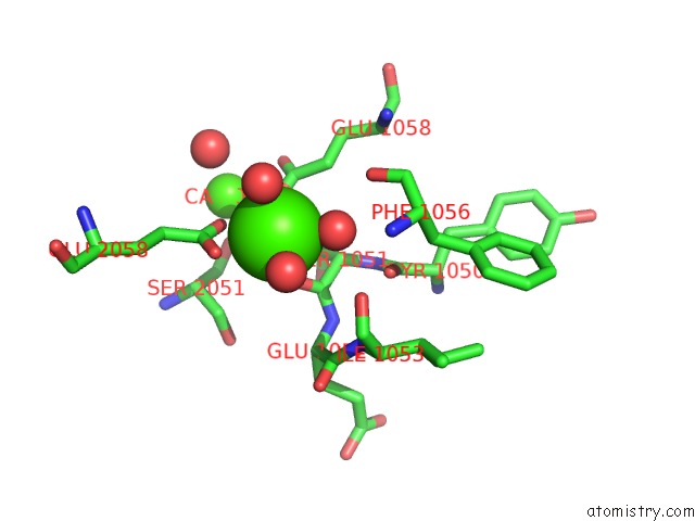
Mono view
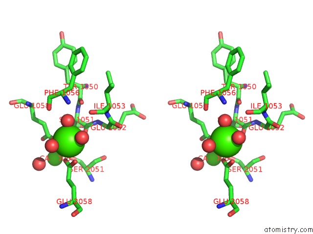
Stereo pair view
Mono view
Stereo pair view
A full contact list of Calcium with other atoms in the Ca binding
site number 2 of Crystal Structure of Calf Spleen Purine Nucleoside Phosphorylase in A New Space Group with Full Trimer in the Asymmetric Unit within 5.0Å range:
|
Calcium binding site 3 out of 6 in 1lv8
Go back to
Calcium binding site 3 out
of 6 in the Crystal Structure of Calf Spleen Purine Nucleoside Phosphorylase in A New Space Group with Full Trimer in the Asymmetric Unit
Mono view
Stereo pair view
Mono view
Stereo pair view
A full contact list of Calcium with other atoms in the Ca binding
site number 3 of Crystal Structure of Calf Spleen Purine Nucleoside Phosphorylase in A New Space Group with Full Trimer in the Asymmetric Unit within 5.0Å range:
|
Calcium binding site 4 out of 6 in 1lv8
Go back to
Calcium binding site 4 out
of 6 in the Crystal Structure of Calf Spleen Purine Nucleoside Phosphorylase in A New Space Group with Full Trimer in the Asymmetric Unit
Mono view
Stereo pair view
Mono view
Stereo pair view
A full contact list of Calcium with other atoms in the Ca binding
site number 4 of Crystal Structure of Calf Spleen Purine Nucleoside Phosphorylase in A New Space Group with Full Trimer in the Asymmetric Unit within 5.0Å range:
|
Calcium binding site 5 out of 6 in 1lv8
Go back to
Calcium binding site 5 out
of 6 in the Crystal Structure of Calf Spleen Purine Nucleoside Phosphorylase in A New Space Group with Full Trimer in the Asymmetric Unit
Mono view
Stereo pair view
Mono view
Stereo pair view
A full contact list of Calcium with other atoms in the Ca binding
site number 5 of Crystal Structure of Calf Spleen Purine Nucleoside Phosphorylase in A New Space Group with Full Trimer in the Asymmetric Unit within 5.0Å range:
|
Calcium binding site 6 out of 6 in 1lv8
Go back to
Calcium binding site 6 out
of 6 in the Crystal Structure of Calf Spleen Purine Nucleoside Phosphorylase in A New Space Group with Full Trimer in the Asymmetric Unit
Mono view
Stereo pair view
Mono view
Stereo pair view
A full contact list of Calcium with other atoms in the Ca binding
site number 6 of Crystal Structure of Calf Spleen Purine Nucleoside Phosphorylase in A New Space Group with Full Trimer in the Asymmetric Unit within 5.0Å range:
|
Reference:
A.Bzowska,
G.Koellner,
B.Wielgus-Kutrowska,
A.Stroh,
G.Raszewski,
A.Holy,
T.Steiner,
J.Frank.
Crystal Structure of Calf Spleen Purine Nucleoside Phosphorylase with Two Full Trimers in the Asymmetric Unit: Important Implications For the Mechanism of Catalysis J.Mol.Biol. V. 342 1015 2004.
ISSN: ISSN 0022-2836
PubMed: 15342253
DOI: 10.1016/J.JMB.2004.07.017
Page generated: Mon Jul 7 17:00:27 2025
ISSN: ISSN 0022-2836
PubMed: 15342253
DOI: 10.1016/J.JMB.2004.07.017
Last articles
I in 5IQYI in 5JTB
I in 5IJQ
I in 5JRV
I in 5JGP
I in 5IO8
I in 5IJS
I in 5IJW
I in 5GZH
I in 5IE1