Calcium »
PDB 1lu1-1m8v »
1m57 »
Calcium in PDB 1m57: Structure of Cytochrome C Oxidase From Rhodobacter Sphaeroides (Eq(I-286) Mutant))
Enzymatic activity of Structure of Cytochrome C Oxidase From Rhodobacter Sphaeroides (Eq(I-286) Mutant))
All present enzymatic activity of Structure of Cytochrome C Oxidase From Rhodobacter Sphaeroides (Eq(I-286) Mutant)):
1.9.3.1;
1.9.3.1;
Protein crystallography data
The structure of Structure of Cytochrome C Oxidase From Rhodobacter Sphaeroides (Eq(I-286) Mutant)), PDB code: 1m57
was solved by
M.Svensson-Ek,
J.Abramson,
G.Larsson,
S.Tornroth,
P.Brezezinski,
S.Iwata,
with X-Ray Crystallography technique. A brief refinement statistics is given in the table below:
| Resolution Low / High (Å) | 4.00 / 3.00 |
| Space group | H 3 |
| Cell size a, b, c (Å), α, β, γ (°) | 340.720, 340.720, 89.760, 90.00, 90.00, 120.00 |
| R / Rfree (%) | 29.3 / 32.9 |
Other elements in 1m57:
The structure of Structure of Cytochrome C Oxidase From Rhodobacter Sphaeroides (Eq(I-286) Mutant)) also contains other interesting chemical elements:
| Magnesium | (Mg) | 2 atoms |
| Iron | (Fe) | 4 atoms |
| Copper | (Cu) | 6 atoms |
Calcium Binding Sites:
The binding sites of Calcium atom in the Structure of Cytochrome C Oxidase From Rhodobacter Sphaeroides (Eq(I-286) Mutant))
(pdb code 1m57). This binding sites where shown within
5.0 Angstroms radius around Calcium atom.
In total 2 binding sites of Calcium where determined in the Structure of Cytochrome C Oxidase From Rhodobacter Sphaeroides (Eq(I-286) Mutant)), PDB code: 1m57:
Jump to Calcium binding site number: 1; 2;
In total 2 binding sites of Calcium where determined in the Structure of Cytochrome C Oxidase From Rhodobacter Sphaeroides (Eq(I-286) Mutant)), PDB code: 1m57:
Jump to Calcium binding site number: 1; 2;
Calcium binding site 1 out of 2 in 1m57
Go back to
Calcium binding site 1 out
of 2 in the Structure of Cytochrome C Oxidase From Rhodobacter Sphaeroides (Eq(I-286) Mutant))
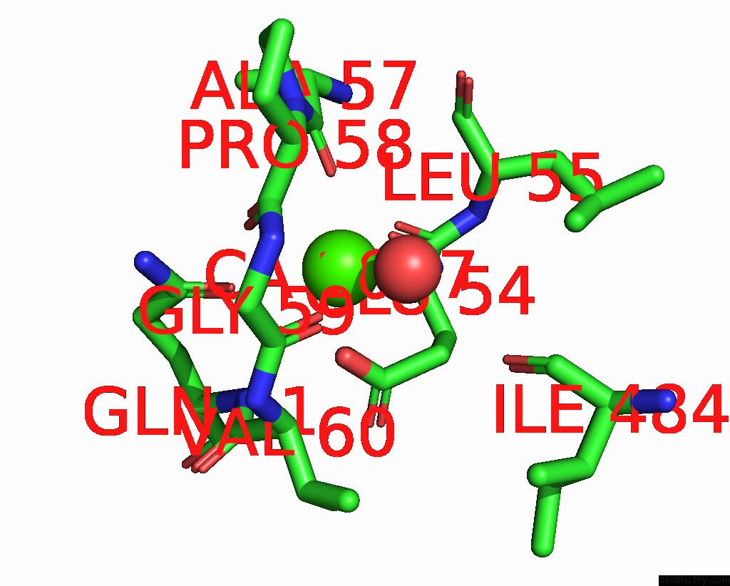
Mono view
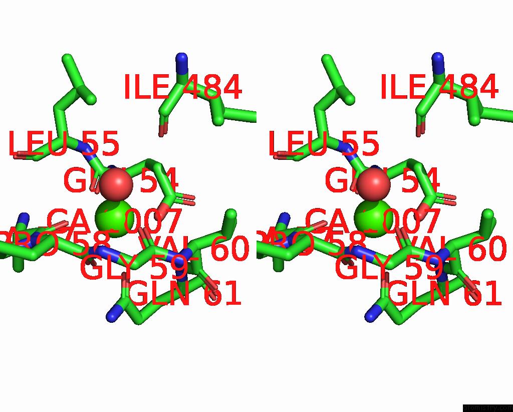
Stereo pair view
Mono view
Stereo pair view
A full contact list of Calcium with other atoms in the Ca binding
site number 1 of Structure of Cytochrome C Oxidase From Rhodobacter Sphaeroides (Eq(I-286) Mutant)) within 5.0Å range:
|
Calcium binding site 2 out of 2 in 1m57
Go back to
Calcium binding site 2 out
of 2 in the Structure of Cytochrome C Oxidase From Rhodobacter Sphaeroides (Eq(I-286) Mutant))
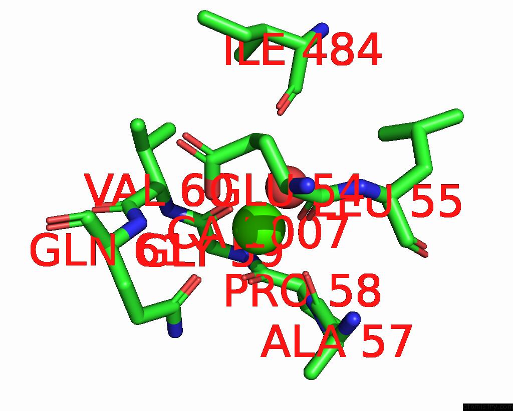
Mono view
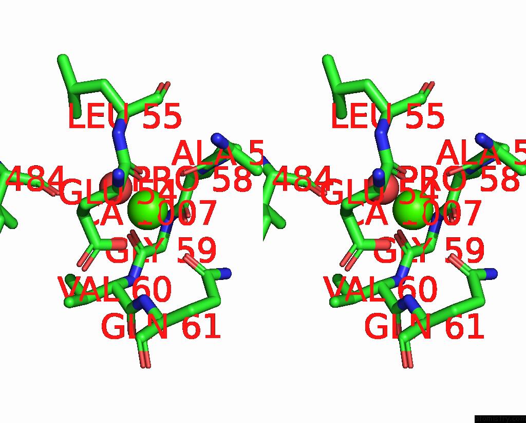
Stereo pair view
Mono view
Stereo pair view
A full contact list of Calcium with other atoms in the Ca binding
site number 2 of Structure of Cytochrome C Oxidase From Rhodobacter Sphaeroides (Eq(I-286) Mutant)) within 5.0Å range:
|
Reference:
M.Svensson-Ek,
J.Abramson,
G.Larsson,
S.Tornroth,
P.Brzezinski,
S.Iwata.
The X-Ray Crystal Structures of Wild-Type and Eq(I-286) Mutant Cytochrome C Oxidases From Rhodobacter Sphaeroides. J.Mol.Biol. V. 321 329 2002.
ISSN: ISSN 0022-2836
PubMed: 12144789
DOI: 10.1016/S0022-2836(02)00619-8
Page generated: Mon Jul 7 17:07:12 2025
ISSN: ISSN 0022-2836
PubMed: 12144789
DOI: 10.1016/S0022-2836(02)00619-8
Last articles
I in 2AK4I in 2ARL
I in 2ANX
I in 2AQW
I in 2AF6
I in 2ANV
I in 2A0N
I in 1XC6
I in 1ZVV
I in 1Z7J