Calcium »
PDB 1lu1-1m8v »
1m8t »
Calcium in PDB 1m8t: Structure of An Acidic Phospholipase A2 From the Venom of Ophiophagus Hannah at 2.1 Resolution From A Hemihedrally Twinned Crystal Form
Enzymatic activity of Structure of An Acidic Phospholipase A2 From the Venom of Ophiophagus Hannah at 2.1 Resolution From A Hemihedrally Twinned Crystal Form
All present enzymatic activity of Structure of An Acidic Phospholipase A2 From the Venom of Ophiophagus Hannah at 2.1 Resolution From A Hemihedrally Twinned Crystal Form:
3.1.1.4;
3.1.1.4;
Protein crystallography data
The structure of Structure of An Acidic Phospholipase A2 From the Venom of Ophiophagus Hannah at 2.1 Resolution From A Hemihedrally Twinned Crystal Form, PDB code: 1m8t
was solved by
S.Xu,
L.Gu,
Q.Wang,
Y.Shu,
Z.Lin,
with X-Ray Crystallography technique. A brief refinement statistics is given in the table below:
| Resolution Low / High (Å) | 16.99 / 2.10 |
| Space group | P 63 |
| Cell size a, b, c (Å), α, β, γ (°) | 98.060, 98.060, 132.390, 90.00, 90.00, 120.00 |
| R / Rfree (%) | 19.2 / 21.3 |
Calcium Binding Sites:
The binding sites of Calcium atom in the Structure of An Acidic Phospholipase A2 From the Venom of Ophiophagus Hannah at 2.1 Resolution From A Hemihedrally Twinned Crystal Form
(pdb code 1m8t). This binding sites where shown within
5.0 Angstroms radius around Calcium atom.
In total 6 binding sites of Calcium where determined in the Structure of An Acidic Phospholipase A2 From the Venom of Ophiophagus Hannah at 2.1 Resolution From A Hemihedrally Twinned Crystal Form, PDB code: 1m8t:
Jump to Calcium binding site number: 1; 2; 3; 4; 5; 6;
In total 6 binding sites of Calcium where determined in the Structure of An Acidic Phospholipase A2 From the Venom of Ophiophagus Hannah at 2.1 Resolution From A Hemihedrally Twinned Crystal Form, PDB code: 1m8t:
Jump to Calcium binding site number: 1; 2; 3; 4; 5; 6;
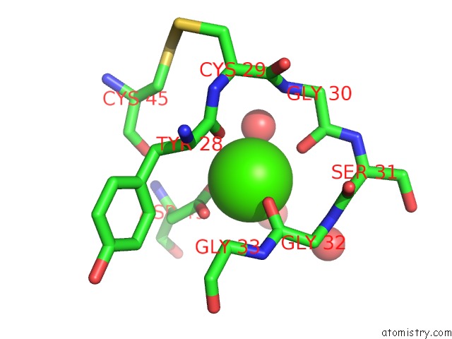
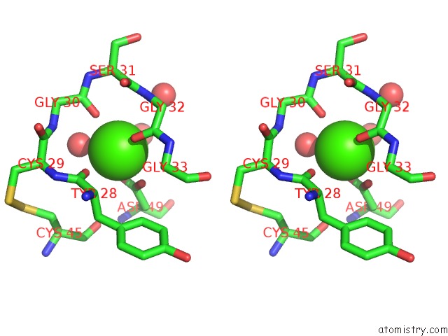
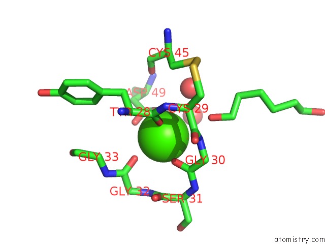
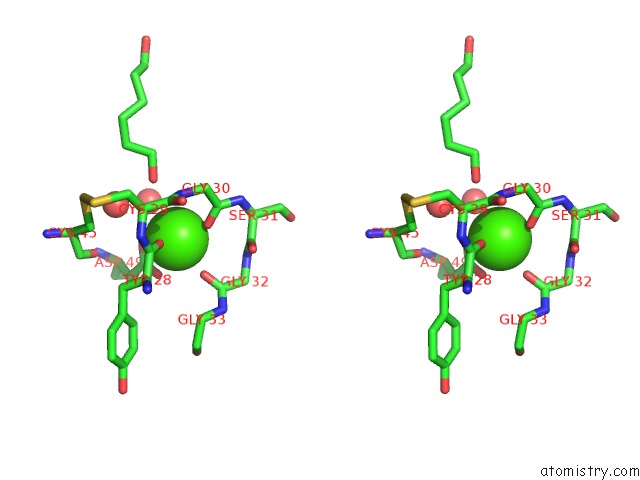
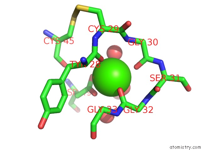
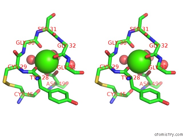
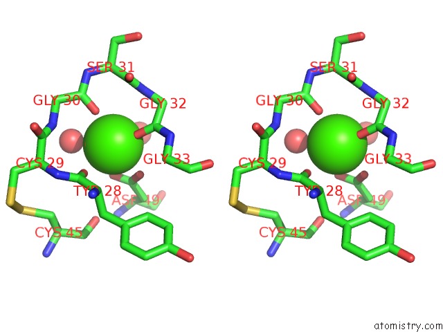
Calcium binding site 1 out of 6 in 1m8t
Go back to
Calcium binding site 1 out
of 6 in the Structure of An Acidic Phospholipase A2 From the Venom of Ophiophagus Hannah at 2.1 Resolution From A Hemihedrally Twinned Crystal Form
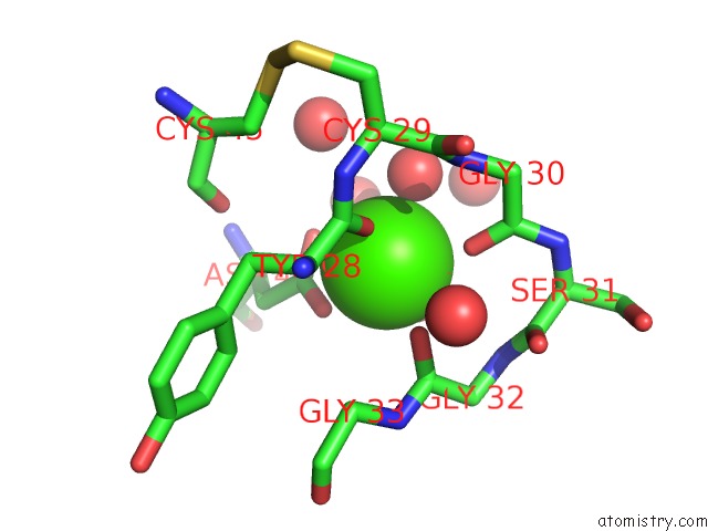
Mono view
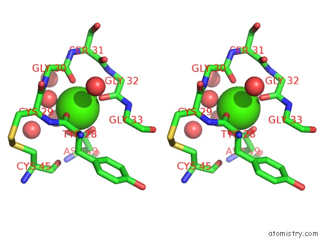
Stereo pair view
Mono view
Stereo pair view
A full contact list of Calcium with other atoms in the Ca binding
site number 1 of Structure of An Acidic Phospholipase A2 From the Venom of Ophiophagus Hannah at 2.1 Resolution From A Hemihedrally Twinned Crystal Form within 5.0Å range:
|
Calcium binding site 2 out of 6 in 1m8t
Go back to
Calcium binding site 2 out
of 6 in the Structure of An Acidic Phospholipase A2 From the Venom of Ophiophagus Hannah at 2.1 Resolution From A Hemihedrally Twinned Crystal Form
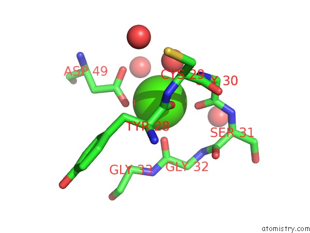
Mono view
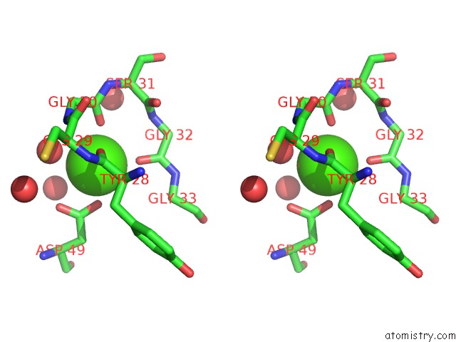
Stereo pair view
Mono view
Stereo pair view
A full contact list of Calcium with other atoms in the Ca binding
site number 2 of Structure of An Acidic Phospholipase A2 From the Venom of Ophiophagus Hannah at 2.1 Resolution From A Hemihedrally Twinned Crystal Form within 5.0Å range:
|
Calcium binding site 3 out of 6 in 1m8t
Go back to
Calcium binding site 3 out
of 6 in the Structure of An Acidic Phospholipase A2 From the Venom of Ophiophagus Hannah at 2.1 Resolution From A Hemihedrally Twinned Crystal Form
Mono view
Stereo pair view
Mono view
Stereo pair view
A full contact list of Calcium with other atoms in the Ca binding
site number 3 of Structure of An Acidic Phospholipase A2 From the Venom of Ophiophagus Hannah at 2.1 Resolution From A Hemihedrally Twinned Crystal Form within 5.0Å range:
|
Calcium binding site 4 out of 6 in 1m8t
Go back to
Calcium binding site 4 out
of 6 in the Structure of An Acidic Phospholipase A2 From the Venom of Ophiophagus Hannah at 2.1 Resolution From A Hemihedrally Twinned Crystal Form
Mono view
Stereo pair view
Mono view
Stereo pair view
A full contact list of Calcium with other atoms in the Ca binding
site number 4 of Structure of An Acidic Phospholipase A2 From the Venom of Ophiophagus Hannah at 2.1 Resolution From A Hemihedrally Twinned Crystal Form within 5.0Å range:
|
Calcium binding site 5 out of 6 in 1m8t
Go back to
Calcium binding site 5 out
of 6 in the Structure of An Acidic Phospholipase A2 From the Venom of Ophiophagus Hannah at 2.1 Resolution From A Hemihedrally Twinned Crystal Form
Mono view
Stereo pair view
Mono view
Stereo pair view
A full contact list of Calcium with other atoms in the Ca binding
site number 5 of Structure of An Acidic Phospholipase A2 From the Venom of Ophiophagus Hannah at 2.1 Resolution From A Hemihedrally Twinned Crystal Form within 5.0Å range:
|
Calcium binding site 6 out of 6 in 1m8t
Go back to
Calcium binding site 6 out
of 6 in the Structure of An Acidic Phospholipase A2 From the Venom of Ophiophagus Hannah at 2.1 Resolution From A Hemihedrally Twinned Crystal Form

Mono view
Stereo pair view
Mono view
Stereo pair view
A full contact list of Calcium with other atoms in the Ca binding
site number 6 of Structure of An Acidic Phospholipase A2 From the Venom of Ophiophagus Hannah at 2.1 Resolution From A Hemihedrally Twinned Crystal Form within 5.0Å range:
|
Reference:
S.Xu,
L.Gu,
Q.Wang,
Y.Shu,
S.Song,
Z.Lin.
Structure of A King Cobra Phospholipase A2 Determined From A Hemihedrally Twinned Crystal. Acta Crystallogr.,Sect.D V. 59 1574 2003.
ISSN: ISSN 0907-4449
PubMed: 12925787
DOI: 10.1107/S0907444903014598
Page generated: Mon Jul 7 17:14:23 2025
ISSN: ISSN 0907-4449
PubMed: 12925787
DOI: 10.1107/S0907444903014598
Last articles
I in 6WYQI in 6WOK
I in 6WNY
I in 6W9D
I in 6WC8
I in 6WE7
I in 6W35
I in 6W0U
I in 6W42
I in 6W2C