Calcium »
PDB 1lu1-1m8v »
1m8v »
Calcium in PDB 1m8v: Structure of Pyrococcus Abyssii Sm Protein in Complex with A Uridine Heptamer
Protein crystallography data
The structure of Structure of Pyrococcus Abyssii Sm Protein in Complex with A Uridine Heptamer, PDB code: 1m8v
was solved by
S.Thore,
C.Mayer,
C.Sauter,
S.Weeks,
D.Suck,
with X-Ray Crystallography technique. A brief refinement statistics is given in the table below:
| Resolution Low / High (Å) | 30.00 / 2.60 |
| Space group | P 1 |
| Cell size a, b, c (Å), α, β, γ (°) | 68.000, 68.000, 84.800, 105.00, 108.80, 100.00 |
| R / Rfree (%) | 21.2 / 28.2 |
Calcium Binding Sites:
The binding sites of Calcium atom in the Structure of Pyrococcus Abyssii Sm Protein in Complex with A Uridine Heptamer
(pdb code 1m8v). This binding sites where shown within
5.0 Angstroms radius around Calcium atom.
In total 7 binding sites of Calcium where determined in the Structure of Pyrococcus Abyssii Sm Protein in Complex with A Uridine Heptamer, PDB code: 1m8v:
Jump to Calcium binding site number: 1; 2; 3; 4; 5; 6; 7;
In total 7 binding sites of Calcium where determined in the Structure of Pyrococcus Abyssii Sm Protein in Complex with A Uridine Heptamer, PDB code: 1m8v:
Jump to Calcium binding site number: 1; 2; 3; 4; 5; 6; 7;
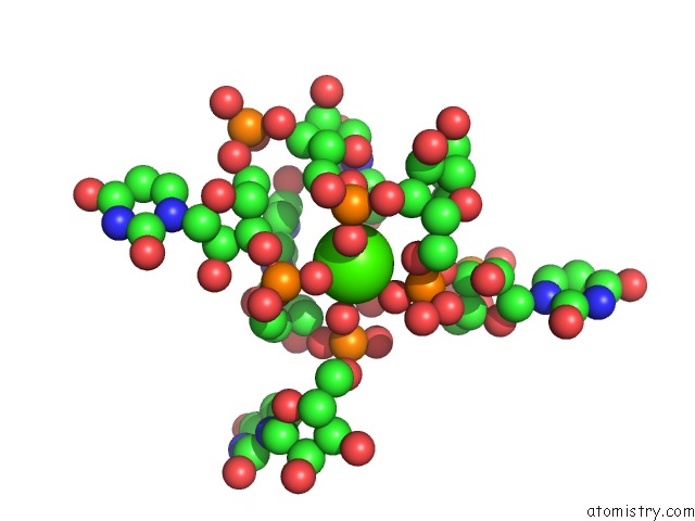
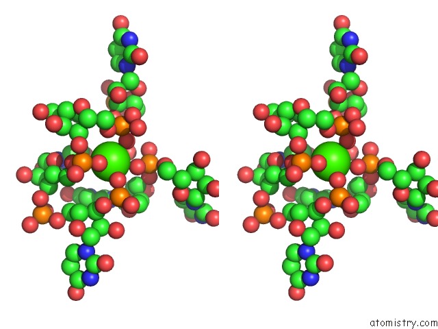
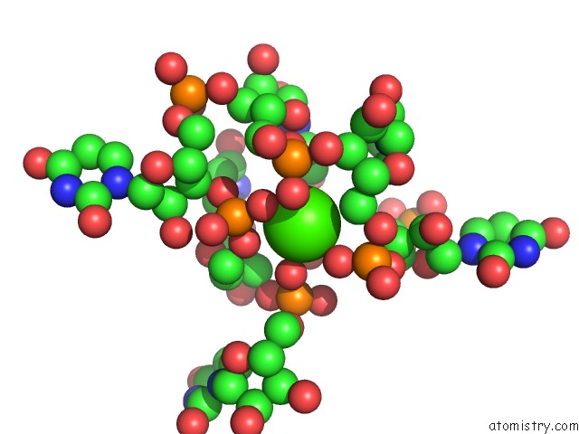
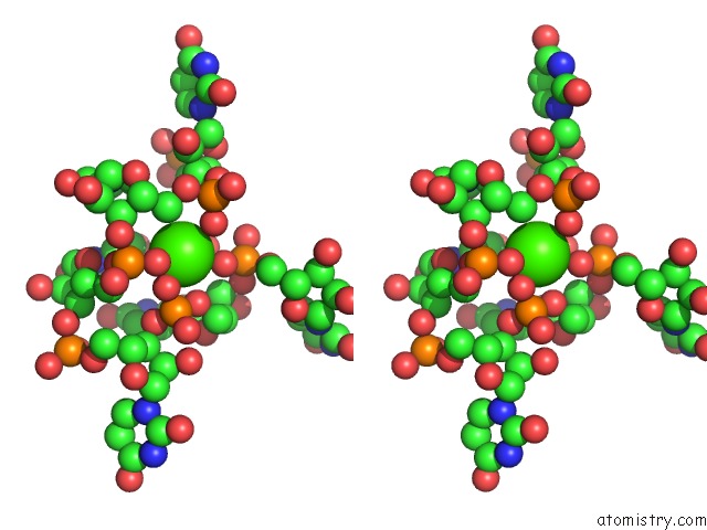
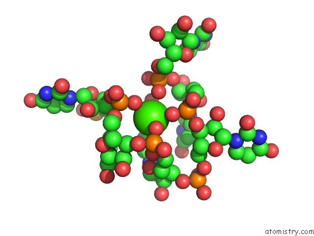
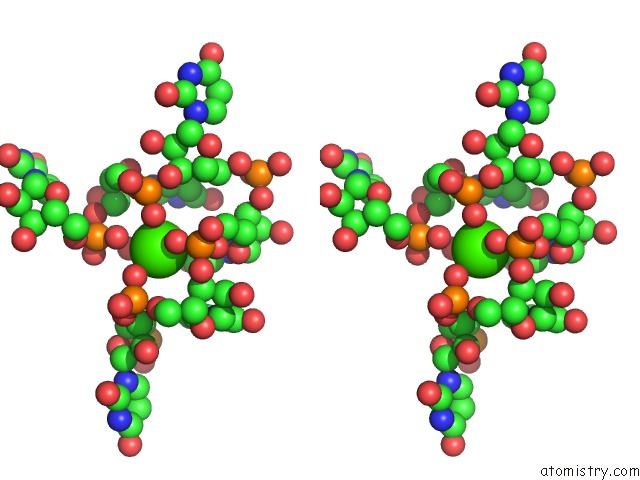
Calcium binding site 1 out of 7 in 1m8v
Go back to
Calcium binding site 1 out
of 7 in the Structure of Pyrococcus Abyssii Sm Protein in Complex with A Uridine Heptamer
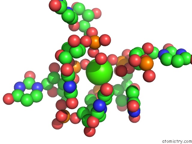
Mono view
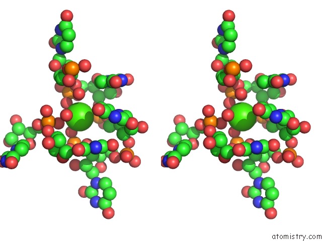
Stereo pair view
Mono view
Stereo pair view
A full contact list of Calcium with other atoms in the Ca binding
site number 1 of Structure of Pyrococcus Abyssii Sm Protein in Complex with A Uridine Heptamer within 5.0Å range:
|
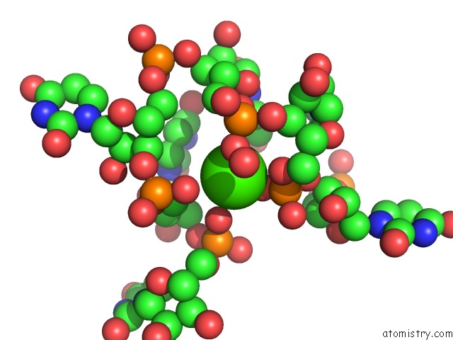
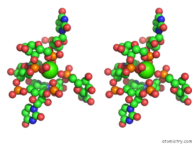
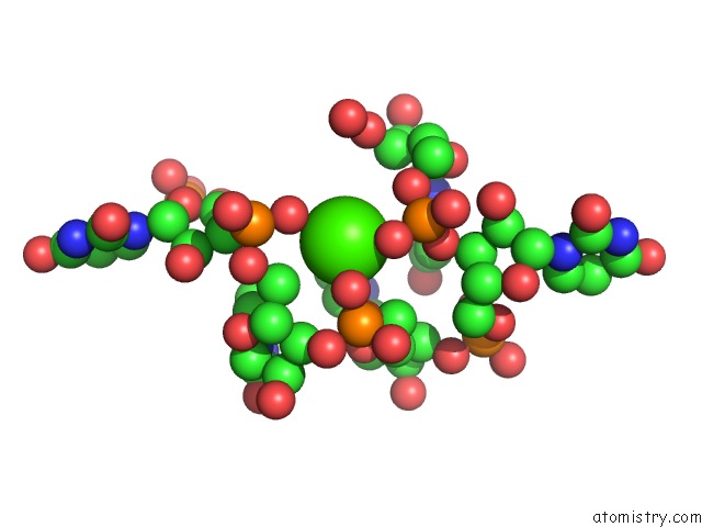
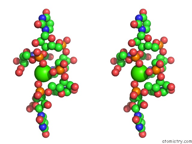
Calcium binding site 2 out of 7 in 1m8v
Go back to
Calcium binding site 2 out
of 7 in the Structure of Pyrococcus Abyssii Sm Protein in Complex with A Uridine Heptamer
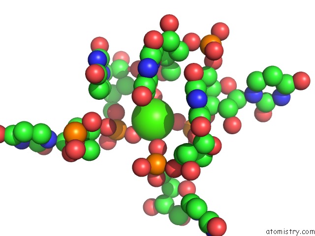
Mono view
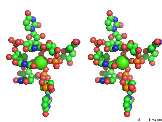
Stereo pair view
Mono view
Stereo pair view
A full contact list of Calcium with other atoms in the Ca binding
site number 2 of Structure of Pyrococcus Abyssii Sm Protein in Complex with A Uridine Heptamer within 5.0Å range:
|
Calcium binding site 3 out of 7 in 1m8v
Go back to
Calcium binding site 3 out
of 7 in the Structure of Pyrococcus Abyssii Sm Protein in Complex with A Uridine Heptamer
Mono view
Stereo pair view
Mono view
Stereo pair view
A full contact list of Calcium with other atoms in the Ca binding
site number 3 of Structure of Pyrococcus Abyssii Sm Protein in Complex with A Uridine Heptamer within 5.0Å range:
|
Calcium binding site 4 out of 7 in 1m8v
Go back to
Calcium binding site 4 out
of 7 in the Structure of Pyrococcus Abyssii Sm Protein in Complex with A Uridine Heptamer
Mono view
Stereo pair view
Mono view
Stereo pair view
A full contact list of Calcium with other atoms in the Ca binding
site number 4 of Structure of Pyrococcus Abyssii Sm Protein in Complex with A Uridine Heptamer within 5.0Å range:
|
Calcium binding site 5 out of 7 in 1m8v
Go back to
Calcium binding site 5 out
of 7 in the Structure of Pyrococcus Abyssii Sm Protein in Complex with A Uridine Heptamer
Mono view
Stereo pair view
Mono view
Stereo pair view
A full contact list of Calcium with other atoms in the Ca binding
site number 5 of Structure of Pyrococcus Abyssii Sm Protein in Complex with A Uridine Heptamer within 5.0Å range:
|
Calcium binding site 6 out of 7 in 1m8v
Go back to
Calcium binding site 6 out
of 7 in the Structure of Pyrococcus Abyssii Sm Protein in Complex with A Uridine Heptamer
Mono view
Stereo pair view
Mono view
Stereo pair view
A full contact list of Calcium with other atoms in the Ca binding
site number 6 of Structure of Pyrococcus Abyssii Sm Protein in Complex with A Uridine Heptamer within 5.0Å range:
|
Calcium binding site 7 out of 7 in 1m8v
Go back to
Calcium binding site 7 out
of 7 in the Structure of Pyrococcus Abyssii Sm Protein in Complex with A Uridine Heptamer
Mono view
Stereo pair view
Mono view
Stereo pair view
A full contact list of Calcium with other atoms in the Ca binding
site number 7 of Structure of Pyrococcus Abyssii Sm Protein in Complex with A Uridine Heptamer within 5.0Å range:
|
Reference:
S.Thore,
C.Mayer,
C.Sauter,
S.Weeks,
D.Suck.
Crystal Structure of Pyrococcus Abyssii Sm Core and Its Complex with Rna: Common Features of Rna-Binding in Archaea and Eukarya J.Biol.Chem. V. 278 1239 2003.
ISSN: ISSN 0021-9258
PubMed: 12409299
DOI: 10.1074/JBC.M207685200
Page generated: Mon Jul 7 17:15:16 2025
ISSN: ISSN 0021-9258
PubMed: 12409299
DOI: 10.1074/JBC.M207685200
Last articles
I in 6W35I in 6W0U
I in 6W42
I in 6W2C
I in 6UIO
I in 6VX2
I in 6VPE
I in 6VU4
I in 6VQS
I in 6VRL