Calcium »
PDB 1mts-1n7d »
1mve »
Calcium in PDB 1mve: Crystal Structure of A Natural Circularly-Permutated Jellyroll Protein: 1,3-1,4-Beta-D-Glucanase From Fibrobacter Succinogenes
Enzymatic activity of Crystal Structure of A Natural Circularly-Permutated Jellyroll Protein: 1,3-1,4-Beta-D-Glucanase From Fibrobacter Succinogenes
All present enzymatic activity of Crystal Structure of A Natural Circularly-Permutated Jellyroll Protein: 1,3-1,4-Beta-D-Glucanase From Fibrobacter Succinogenes:
3.2.1.73;
3.2.1.73;
Protein crystallography data
The structure of Crystal Structure of A Natural Circularly-Permutated Jellyroll Protein: 1,3-1,4-Beta-D-Glucanase From Fibrobacter Succinogenes, PDB code: 1mve
was solved by
L.-C.Tsai,
L.-F.Shyur,
S.-H.Lee,
S.-S.Lin,
H.S.Yuan,
with X-Ray Crystallography technique. A brief refinement statistics is given in the table below:
| Resolution Low / High (Å) | 32.93 / 1.70 |
| Space group | P 21 21 21 |
| Cell size a, b, c (Å), α, β, γ (°) | 40.850, 73.350, 73.710, 90.00, 90.00, 90.00 |
| R / Rfree (%) | 19.2 / 23.7 |
Calcium Binding Sites:
The binding sites of Calcium atom in the Crystal Structure of A Natural Circularly-Permutated Jellyroll Protein: 1,3-1,4-Beta-D-Glucanase From Fibrobacter Succinogenes
(pdb code 1mve). This binding sites where shown within
5.0 Angstroms radius around Calcium atom.
In total only one binding site of Calcium was determined in the Crystal Structure of A Natural Circularly-Permutated Jellyroll Protein: 1,3-1,4-Beta-D-Glucanase From Fibrobacter Succinogenes, PDB code: 1mve:
In total only one binding site of Calcium was determined in the Crystal Structure of A Natural Circularly-Permutated Jellyroll Protein: 1,3-1,4-Beta-D-Glucanase From Fibrobacter Succinogenes, PDB code: 1mve:
Calcium binding site 1 out of 1 in 1mve
Go back to
Calcium binding site 1 out
of 1 in the Crystal Structure of A Natural Circularly-Permutated Jellyroll Protein: 1,3-1,4-Beta-D-Glucanase From Fibrobacter Succinogenes
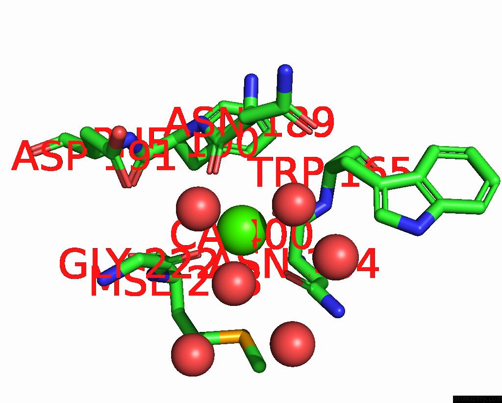
Mono view
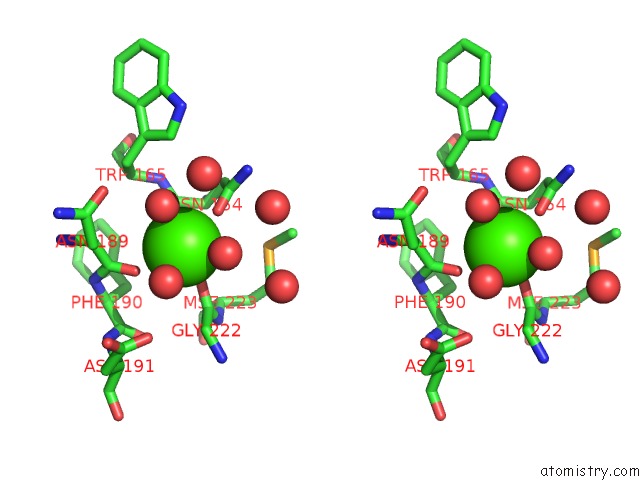
Stereo pair view
Mono view
Stereo pair view
A full contact list of Calcium with other atoms in the Ca binding
site number 1 of Crystal Structure of A Natural Circularly-Permutated Jellyroll Protein: 1,3-1,4-Beta-D-Glucanase From Fibrobacter Succinogenes within 5.0Å range:
|
Reference:
L.C.Tsai,
L.F.Shyur,
S.H.Lee,
S.S.Lin,
H.S.Yuan.
Crystal Structure of A Natural Circularly Permuted Jellyroll Protein: 1,3-1,4-Beta-D-Glucanase From Fibrobacter Succinogenes. J.Mol.Biol. V. 330 607 2003.
ISSN: ISSN 0022-2836
PubMed: 12842475
DOI: 10.1016/S0022-2836(03)00630-2
Page generated: Mon Jul 7 17:22:20 2025
ISSN: ISSN 0022-2836
PubMed: 12842475
DOI: 10.1016/S0022-2836(03)00630-2
Last articles
Mg in 3A14Mg in 3A11
Mg in 3A10
Mg in 3A0U
Mg in 3A0H
Mg in 3A0B
Mg in 364D
Mg in 3A0T
Mg in 357D
Mg in 3A06