Calcium »
PDB 1mts-1n7d »
1n29 »
Calcium in PDB 1n29: Crystal Structure of the N1A Mutant of Human Group Iia Phospholipase A2
Enzymatic activity of Crystal Structure of the N1A Mutant of Human Group Iia Phospholipase A2
All present enzymatic activity of Crystal Structure of the N1A Mutant of Human Group Iia Phospholipase A2:
3.1.1.4;
3.1.1.4;
Protein crystallography data
The structure of Crystal Structure of the N1A Mutant of Human Group Iia Phospholipase A2, PDB code: 1n29
was solved by
S.H.Edwards,
D.Thompson,
S.F.Baker,
S.P.Wood,
D.C.Wilton,
with X-Ray Crystallography technique. A brief refinement statistics is given in the table below:
| Resolution Low / High (Å) | 15.00 / 2.60 |
| Space group | P 61 2 2 |
| Cell size a, b, c (Å), α, β, γ (°) | 74.790, 74.790, 88.891, 90.00, 90.00, 120.00 |
| R / Rfree (%) | 24.2 / 32 |
Calcium Binding Sites:
The binding sites of Calcium atom in the Crystal Structure of the N1A Mutant of Human Group Iia Phospholipase A2
(pdb code 1n29). This binding sites where shown within
5.0 Angstroms radius around Calcium atom.
In total 2 binding sites of Calcium where determined in the Crystal Structure of the N1A Mutant of Human Group Iia Phospholipase A2, PDB code: 1n29:
Jump to Calcium binding site number: 1; 2;
In total 2 binding sites of Calcium where determined in the Crystal Structure of the N1A Mutant of Human Group Iia Phospholipase A2, PDB code: 1n29:
Jump to Calcium binding site number: 1; 2;
Calcium binding site 1 out of 2 in 1n29
Go back to
Calcium binding site 1 out
of 2 in the Crystal Structure of the N1A Mutant of Human Group Iia Phospholipase A2
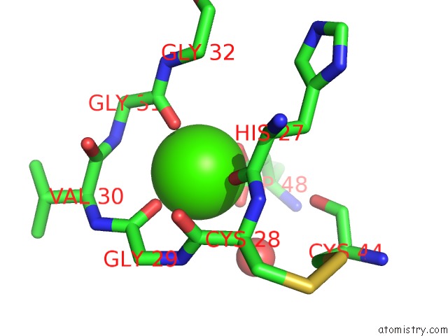
Mono view
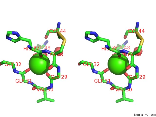
Stereo pair view
Mono view
Stereo pair view
A full contact list of Calcium with other atoms in the Ca binding
site number 1 of Crystal Structure of the N1A Mutant of Human Group Iia Phospholipase A2 within 5.0Å range:
|
Calcium binding site 2 out of 2 in 1n29
Go back to
Calcium binding site 2 out
of 2 in the Crystal Structure of the N1A Mutant of Human Group Iia Phospholipase A2

Mono view

Stereo pair view
Mono view
Stereo pair view
A full contact list of Calcium with other atoms in the Ca binding
site number 2 of Crystal Structure of the N1A Mutant of Human Group Iia Phospholipase A2 within 5.0Å range:
|
Reference:
S.H.Edwards,
D.Thompson,
S.F.Baker,
S.P.Wood,
D.C.Wilton.
The Crystal Structure of the H48Q Active Site Mutant of Human Group Iia Secreted Phospholipase A2 at 1.5 A Resolution Provides An Insight Into the Catalytic Mechanism Biochemistry V. 41 15468 2002.
ISSN: ISSN 0006-2960
PubMed: 12501175
DOI: 10.1021/BI020485Z
Page generated: Mon Jul 7 17:24:37 2025
ISSN: ISSN 0006-2960
PubMed: 12501175
DOI: 10.1021/BI020485Z
Last articles
Mg in 3D2JMg in 3D2E
Mg in 3D1R
Mg in 3CXC
Mg in 3CPW
Mg in 3CZJ
Mg in 3D19
Mg in 3CZ4
Mg in 3CZ1
Mg in 3CZ0