Calcium »
PDB 1nmb-1nxc »
1nps »
Calcium in PDB 1nps: Crystal Structure of N-Terminal Domain of Protein S
Protein crystallography data
The structure of Crystal Structure of N-Terminal Domain of Protein S, PDB code: 1nps
was solved by
M.Wenk,
R.Baumgartner,
E.M.Mayer,
R.Huber,
T.A.Holak,
R.Jaenicke,
with X-Ray Crystallography technique. A brief refinement statistics is given in the table below:
| Resolution Low / High (Å) | 20.00 / 1.80 |
| Space group | P 1 21 1 |
| Cell size a, b, c (Å), α, β, γ (°) | 28.360, 37.940, 37.260, 90.00, 105.93, 90.00 |
| R / Rfree (%) | 20 / 23.4 |
Calcium Binding Sites:
The binding sites of Calcium atom in the Crystal Structure of N-Terminal Domain of Protein S
(pdb code 1nps). This binding sites where shown within
5.0 Angstroms radius around Calcium atom.
In total 2 binding sites of Calcium where determined in the Crystal Structure of N-Terminal Domain of Protein S, PDB code: 1nps:
Jump to Calcium binding site number: 1; 2;
In total 2 binding sites of Calcium where determined in the Crystal Structure of N-Terminal Domain of Protein S, PDB code: 1nps:
Jump to Calcium binding site number: 1; 2;
Calcium binding site 1 out of 2 in 1nps
Go back to
Calcium binding site 1 out
of 2 in the Crystal Structure of N-Terminal Domain of Protein S
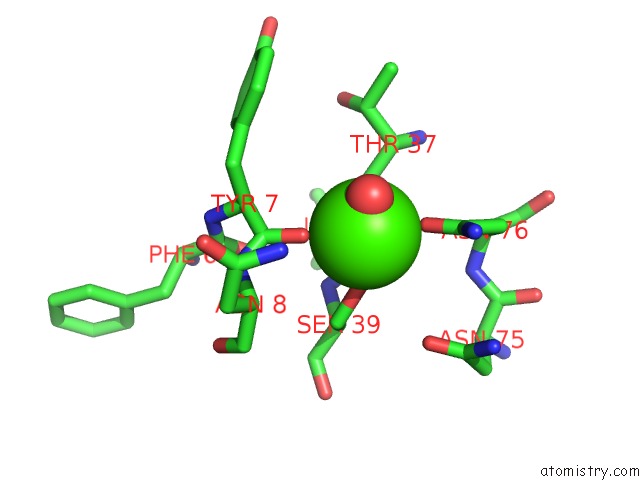
Mono view
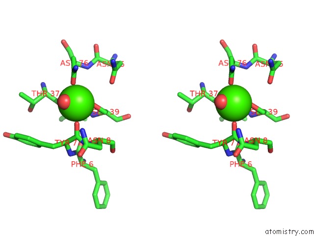
Stereo pair view
Mono view
Stereo pair view
A full contact list of Calcium with other atoms in the Ca binding
site number 1 of Crystal Structure of N-Terminal Domain of Protein S within 5.0Å range:
|
Calcium binding site 2 out of 2 in 1nps
Go back to
Calcium binding site 2 out
of 2 in the Crystal Structure of N-Terminal Domain of Protein S
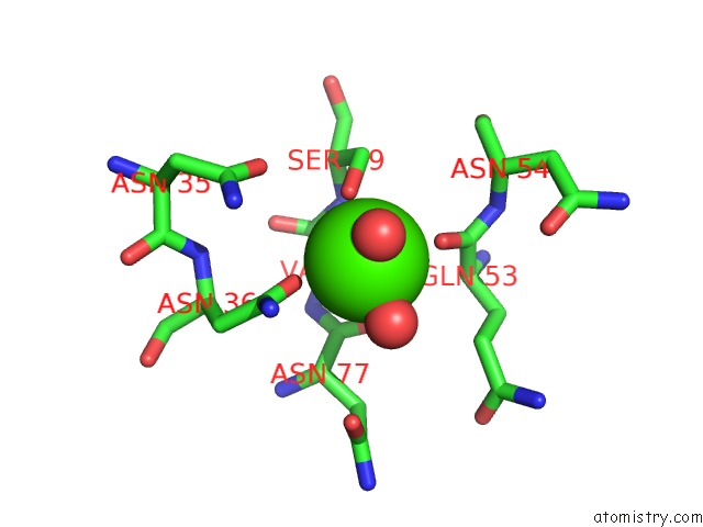
Mono view
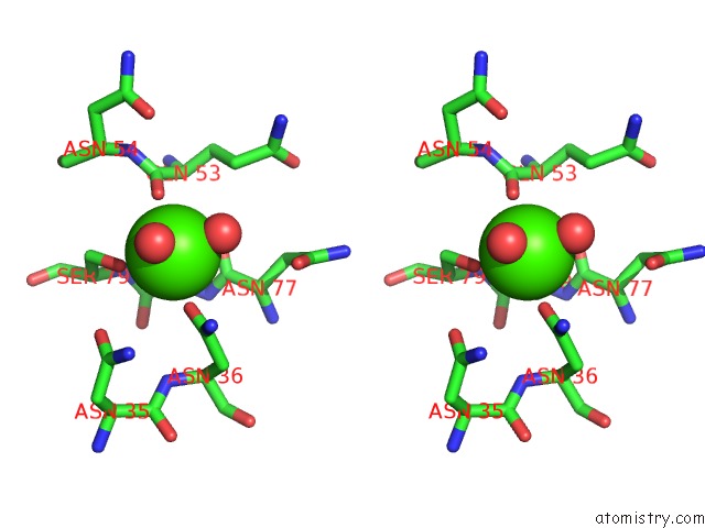
Stereo pair view
Mono view
Stereo pair view
A full contact list of Calcium with other atoms in the Ca binding
site number 2 of Crystal Structure of N-Terminal Domain of Protein S within 5.0Å range:
|
Reference:
M.Wenk,
R.Baumgartner,
T.A.Holak,
R.Huber,
R.Jaenicke,
E.M.Mayr.
The Domains of Protein S From Myxococcus Xanthus: Structure, Stability and Interactions. J.Mol.Biol. V. 286 1533 1999.
ISSN: ISSN 0022-2836
PubMed: 10064714
DOI: 10.1006/JMBI.1999.2582
Page generated: Mon Jul 7 17:38:20 2025
ISSN: ISSN 0022-2836
PubMed: 10064714
DOI: 10.1006/JMBI.1999.2582
Last articles
I in 3UNHI in 3UNF
I in 3UBA
I in 3UFM
I in 3TUR
I in 3UCO
I in 3U8T
I in 3U8R
I in 3U8O
I in 3TGY