Calcium »
PDB 1nya-1o3c »
1o23 »
Calcium in PDB 1o23: Crystal Structure of Lactose Synthase in the Presence of Udp-Glucose
Enzymatic activity of Crystal Structure of Lactose Synthase in the Presence of Udp-Glucose
All present enzymatic activity of Crystal Structure of Lactose Synthase in the Presence of Udp-Glucose:
2.4.1.90;
2.4.1.90;
Protein crystallography data
The structure of Crystal Structure of Lactose Synthase in the Presence of Udp-Glucose, PDB code: 1o23
was solved by
B.Ramakrishnan,
P.S.Shah,
P.K.Qasba,
with X-Ray Crystallography technique. A brief refinement statistics is given in the table below:
| Resolution Low / High (Å) | 19.96 / 2.32 |
| Space group | P 1 21 1 |
| Cell size a, b, c (Å), α, β, γ (°) | 55.545, 99.379, 102.568, 90.00, 104.09, 90.00 |
| R / Rfree (%) | 20.1 / 26.5 |
Other elements in 1o23:
The structure of Crystal Structure of Lactose Synthase in the Presence of Udp-Glucose also contains other interesting chemical elements:
| Manganese | (Mn) | 2 atoms |
Calcium Binding Sites:
The binding sites of Calcium atom in the Crystal Structure of Lactose Synthase in the Presence of Udp-Glucose
(pdb code 1o23). This binding sites where shown within
5.0 Angstroms radius around Calcium atom.
In total 2 binding sites of Calcium where determined in the Crystal Structure of Lactose Synthase in the Presence of Udp-Glucose, PDB code: 1o23:
Jump to Calcium binding site number: 1; 2;
In total 2 binding sites of Calcium where determined in the Crystal Structure of Lactose Synthase in the Presence of Udp-Glucose, PDB code: 1o23:
Jump to Calcium binding site number: 1; 2;
Calcium binding site 1 out of 2 in 1o23
Go back to
Calcium binding site 1 out
of 2 in the Crystal Structure of Lactose Synthase in the Presence of Udp-Glucose
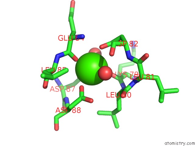
Mono view
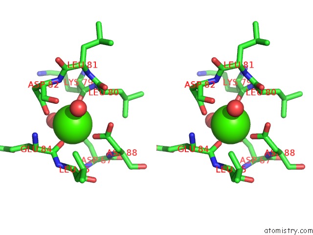
Stereo pair view
Mono view
Stereo pair view
A full contact list of Calcium with other atoms in the Ca binding
site number 1 of Crystal Structure of Lactose Synthase in the Presence of Udp-Glucose within 5.0Å range:
|
Calcium binding site 2 out of 2 in 1o23
Go back to
Calcium binding site 2 out
of 2 in the Crystal Structure of Lactose Synthase in the Presence of Udp-Glucose
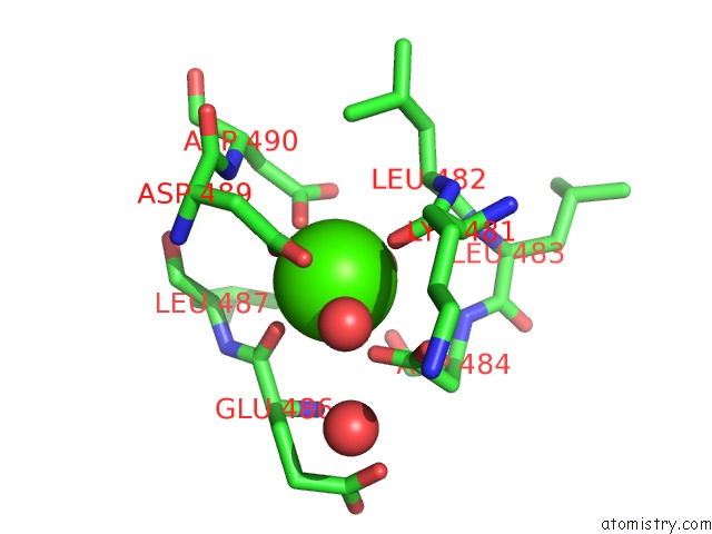
Mono view
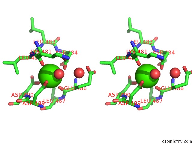
Stereo pair view
Mono view
Stereo pair view
A full contact list of Calcium with other atoms in the Ca binding
site number 2 of Crystal Structure of Lactose Synthase in the Presence of Udp-Glucose within 5.0Å range:
|
Reference:
B.Ramakrishnan,
P.S.Shah,
P.K.Qasba.
Alpha-Lactalbumin (La) Stimulates Milk Beta-1,4-Galactosyltransferase I (Beta 4GAL-T1) to Transfer Glucose From Udp-Glucose to N-Acetylglucosamine. Crystal Structure of Beta 4GAL-T1 X La Complex with Udp-Glc. J.Biol.Chem. V. 276 37665 2001.
ISSN: ISSN 0021-9258
PubMed: 11485999
DOI: 10.1074/JBC.M102458200
Page generated: Mon Jul 7 17:46:03 2025
ISSN: ISSN 0021-9258
PubMed: 11485999
DOI: 10.1074/JBC.M102458200
Last articles
Mg in 1IZLMg in 1JBZ
Mg in 1JBW
Mg in 1JBV
Mg in 1JBK
Mg in 1JAX
Mg in 1JAH
Mg in 1J97
Mg in 1J9J
Mg in 1J8L