Calcium »
PDB 1nya-1o3c »
1o2e »
Calcium in PDB 1o2e: Structure of the Triple Mutant (K53,56,120M) + Anisic Acid Complex of Phospholipase A2
Enzymatic activity of Structure of the Triple Mutant (K53,56,120M) + Anisic Acid Complex of Phospholipase A2
All present enzymatic activity of Structure of the Triple Mutant (K53,56,120M) + Anisic Acid Complex of Phospholipase A2:
3.1.1.4;
3.1.1.4;
Protein crystallography data
The structure of Structure of the Triple Mutant (K53,56,120M) + Anisic Acid Complex of Phospholipase A2, PDB code: 1o2e
was solved by
K.Sekar,
D.Velmurugan,
M.D.Tsai,
with X-Ray Crystallography technique. A brief refinement statistics is given in the table below:
| Resolution Low / High (Å) | 19.80 / 2.60 |
| Space group | P 31 2 1 |
| Cell size a, b, c (Å), α, β, γ (°) | 46.610, 46.610, 102.680, 90.00, 90.00, 120.00 |
| R / Rfree (%) | 18.7 / 24.1 |
Calcium Binding Sites:
The binding sites of Calcium atom in the Structure of the Triple Mutant (K53,56,120M) + Anisic Acid Complex of Phospholipase A2
(pdb code 1o2e). This binding sites where shown within
5.0 Angstroms radius around Calcium atom.
In total only one binding site of Calcium was determined in the Structure of the Triple Mutant (K53,56,120M) + Anisic Acid Complex of Phospholipase A2, PDB code: 1o2e:
In total only one binding site of Calcium was determined in the Structure of the Triple Mutant (K53,56,120M) + Anisic Acid Complex of Phospholipase A2, PDB code: 1o2e:
Calcium binding site 1 out of 1 in 1o2e
Go back to
Calcium binding site 1 out
of 1 in the Structure of the Triple Mutant (K53,56,120M) + Anisic Acid Complex of Phospholipase A2
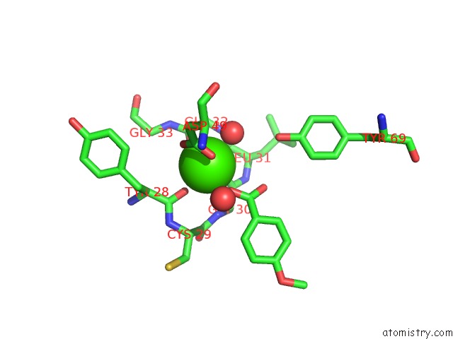
Mono view
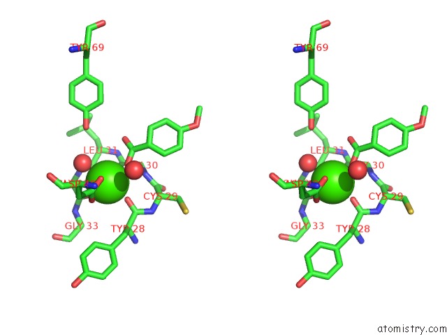
Stereo pair view
Mono view
Stereo pair view
A full contact list of Calcium with other atoms in the Ca binding
site number 1 of Structure of the Triple Mutant (K53,56,120M) + Anisic Acid Complex of Phospholipase A2 within 5.0Å range:
|
Reference:
K.Sekar,
S.Vaijayanthi Mala,
M.Yogavel,
D.Velmurugan,
M.J.Poi,
B.S.Vishwanath,
T.V.Gowda,
A.A.Jeyaprakash,
M.D.Tsai.
Crystal Structures of the Free and Anisic Acid Bound Triple Mutant of Phospholipase A2. J.Mol.Biol. V. 333 367 2003.
ISSN: ISSN 0022-2836
PubMed: 14529623
DOI: 10.1016/J.JMB.2003.08.032
Page generated: Mon Jul 7 17:46:18 2025
ISSN: ISSN 0022-2836
PubMed: 14529623
DOI: 10.1016/J.JMB.2003.08.032
Last articles
Fe in 2YXOFe in 2YRS
Fe in 2YXC
Fe in 2YNM
Fe in 2YVJ
Fe in 2YP1
Fe in 2YU2
Fe in 2YU1
Fe in 2YQB
Fe in 2YOO