Calcium »
PDB 1o3d-1odb »
1o5k »
Calcium in PDB 1o5k: Crystal Structure of Dihydrodipicolinate Synthase (TM1521) From Thermotoga Maritima at 1.80 A Resolution
Enzymatic activity of Crystal Structure of Dihydrodipicolinate Synthase (TM1521) From Thermotoga Maritima at 1.80 A Resolution
All present enzymatic activity of Crystal Structure of Dihydrodipicolinate Synthase (TM1521) From Thermotoga Maritima at 1.80 A Resolution:
4.3.3.7;
4.3.3.7;
Protein crystallography data
The structure of Crystal Structure of Dihydrodipicolinate Synthase (TM1521) From Thermotoga Maritima at 1.80 A Resolution, PDB code: 1o5k
was solved by
Joint Center For Structural Genomics (Jcsg),
with X-Ray Crystallography technique. A brief refinement statistics is given in the table below:
| Resolution Low / High (Å) | 35.61 / 1.80 |
| Space group | C 2 2 21 |
| Cell size a, b, c (Å), α, β, γ (°) | 54.672, 140.768, 155.943, 90.00, 90.00, 90.00 |
| R / Rfree (%) | 13.9 / 18.6 |
Calcium Binding Sites:
The binding sites of Calcium atom in the Crystal Structure of Dihydrodipicolinate Synthase (TM1521) From Thermotoga Maritima at 1.80 A Resolution
(pdb code 1o5k). This binding sites where shown within
5.0 Angstroms radius around Calcium atom.
In total only one binding site of Calcium was determined in the Crystal Structure of Dihydrodipicolinate Synthase (TM1521) From Thermotoga Maritima at 1.80 A Resolution, PDB code: 1o5k:
In total only one binding site of Calcium was determined in the Crystal Structure of Dihydrodipicolinate Synthase (TM1521) From Thermotoga Maritima at 1.80 A Resolution, PDB code: 1o5k:
Calcium binding site 1 out of 1 in 1o5k
Go back to
Calcium binding site 1 out
of 1 in the Crystal Structure of Dihydrodipicolinate Synthase (TM1521) From Thermotoga Maritima at 1.80 A Resolution

Mono view
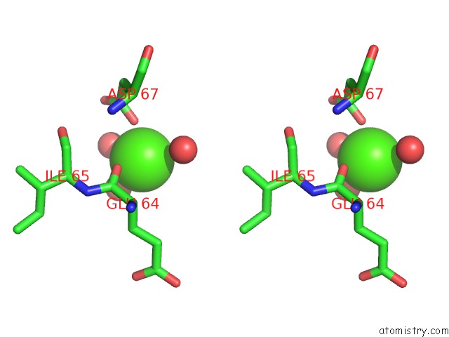
Stereo pair view
Mono view
Stereo pair view
A full contact list of Calcium with other atoms in the Ca binding
site number 1 of Crystal Structure of Dihydrodipicolinate Synthase (TM1521) From Thermotoga Maritima at 1.80 A Resolution within 5.0Å range:
|
Reference:
Joint Center For Structural Genomics (Jcsg),
Joint Center For Structural Genomics (Jcsg).
N/A N/A.
Page generated: Mon Jul 7 17:50:38 2025
Last articles
Mg in 6IMTMg in 6IMO
Mg in 6IMI
Mg in 6IMR
Mg in 6IMD
Mg in 6IME
Mg in 6IMB
Mg in 6IM6
Mg in 6ILT
Mg in 6IKA