Calcium »
PDB 1pig-1px2 »
1pph »
Calcium in PDB 1pph: Geometry of Binding of the Nalpha-Tosylated Piperidides of M-Amidino-, P-Amidino-and P-Guanidino Phenylalanine to Thrombin and Trypsin: X- Ray Crystal Structures of Their Trypsin Complexes and Modeling of Their Thrombin Complexes
Enzymatic activity of Geometry of Binding of the Nalpha-Tosylated Piperidides of M-Amidino-, P-Amidino-and P-Guanidino Phenylalanine to Thrombin and Trypsin: X- Ray Crystal Structures of Their Trypsin Complexes and Modeling of Their Thrombin Complexes
All present enzymatic activity of Geometry of Binding of the Nalpha-Tosylated Piperidides of M-Amidino-, P-Amidino-and P-Guanidino Phenylalanine to Thrombin and Trypsin: X- Ray Crystal Structures of Their Trypsin Complexes and Modeling of Their Thrombin Complexes:
3.4.21.4;
3.4.21.4;
Protein crystallography data
The structure of Geometry of Binding of the Nalpha-Tosylated Piperidides of M-Amidino-, P-Amidino-and P-Guanidino Phenylalanine to Thrombin and Trypsin: X- Ray Crystal Structures of Their Trypsin Complexes and Modeling of Their Thrombin Complexes, PDB code: 1pph
was solved by
W.Bode,
D.Turk,
with X-Ray Crystallography technique. A brief refinement statistics is given in the table below:
| Resolution Low / High (Å) | 8.00 / 1.90 |
| Space group | P 21 21 21 |
| Cell size a, b, c (Å), α, β, γ (°) | 63.510, 69.190, 63.810, 90.00, 90.00, 90.00 |
| R / Rfree (%) | 16.7 / n/a |
Calcium Binding Sites:
The binding sites of Calcium atom in the Geometry of Binding of the Nalpha-Tosylated Piperidides of M-Amidino-, P-Amidino-and P-Guanidino Phenylalanine to Thrombin and Trypsin: X- Ray Crystal Structures of Their Trypsin Complexes and Modeling of Their Thrombin Complexes
(pdb code 1pph). This binding sites where shown within
5.0 Angstroms radius around Calcium atom.
In total only one binding site of Calcium was determined in the Geometry of Binding of the Nalpha-Tosylated Piperidides of M-Amidino-, P-Amidino-and P-Guanidino Phenylalanine to Thrombin and Trypsin: X- Ray Crystal Structures of Their Trypsin Complexes and Modeling of Their Thrombin Complexes, PDB code: 1pph:
In total only one binding site of Calcium was determined in the Geometry of Binding of the Nalpha-Tosylated Piperidides of M-Amidino-, P-Amidino-and P-Guanidino Phenylalanine to Thrombin and Trypsin: X- Ray Crystal Structures of Their Trypsin Complexes and Modeling of Their Thrombin Complexes, PDB code: 1pph:
Calcium binding site 1 out of 1 in 1pph
Go back to
Calcium binding site 1 out
of 1 in the Geometry of Binding of the Nalpha-Tosylated Piperidides of M-Amidino-, P-Amidino-and P-Guanidino Phenylalanine to Thrombin and Trypsin: X- Ray Crystal Structures of Their Trypsin Complexes and Modeling of Their Thrombin Complexes
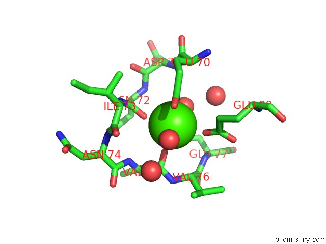
Mono view
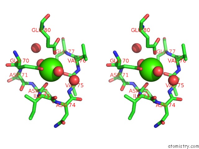
Stereo pair view
Mono view
Stereo pair view
A full contact list of Calcium with other atoms in the Ca binding
site number 1 of Geometry of Binding of the Nalpha-Tosylated Piperidides of M-Amidino-, P-Amidino-and P-Guanidino Phenylalanine to Thrombin and Trypsin: X- Ray Crystal Structures of Their Trypsin Complexes and Modeling of Their Thrombin Complexes within 5.0Å range:
|
Reference:
D.Turk,
J.Sturzebecher,
W.Bode.
Geometry of Binding of the N Alpha-Tosylated Piperidides of M-Amidino-, P-Amidino- and P-Guanidino Phenylalanine to Thrombin and Trypsin. X-Ray Crystal Structures of Their Trypsin Complexes and Modeling of Their Thrombin Complexes. Febs Lett. V. 287 133 1991.
ISSN: ISSN 0014-5793
PubMed: 1879520
DOI: 10.1016/0014-5793(91)80033-Y
Page generated: Mon Jul 7 18:16:49 2025
ISSN: ISSN 0014-5793
PubMed: 1879520
DOI: 10.1016/0014-5793(91)80033-Y
Last articles
Mg in 4DV0Mg in 4DV1
Mg in 4DUZ
Mg in 4DUY
Mg in 4DR7
Mg in 4DR6
Mg in 4DR5
Mg in 4DUX
Mg in 4DUW
Mg in 4DUV