Calcium »
PDB 1qls-1ra1 »
1qos »
Calcium in PDB 1qos: Lectin Uea-II Complexed with Chitobiose
Protein crystallography data
The structure of Lectin Uea-II Complexed with Chitobiose, PDB code: 1qos
was solved by
R.Loris,
H.De Greve,
M.-H.Dao-Thi,
J.Messens,
A.Imberty,
L.Wyns,
with X-Ray Crystallography technique. A brief refinement statistics is given in the table below:
| Resolution Low / High (Å) | 20.00 / 2.95 |
| Space group | P 31 2 1 |
| Cell size a, b, c (Å), α, β, γ (°) | 106.030, 106.030, 87.020, 90.00, 90.00, 120.00 |
| R / Rfree (%) | 18.9 / 22.3 |
Other elements in 1qos:
The structure of Lectin Uea-II Complexed with Chitobiose also contains other interesting chemical elements:
| Manganese | (Mn) | 2 atoms |
Calcium Binding Sites:
The binding sites of Calcium atom in the Lectin Uea-II Complexed with Chitobiose
(pdb code 1qos). This binding sites where shown within
5.0 Angstroms radius around Calcium atom.
In total 2 binding sites of Calcium where determined in the Lectin Uea-II Complexed with Chitobiose, PDB code: 1qos:
Jump to Calcium binding site number: 1; 2;
In total 2 binding sites of Calcium where determined in the Lectin Uea-II Complexed with Chitobiose, PDB code: 1qos:
Jump to Calcium binding site number: 1; 2;
Calcium binding site 1 out of 2 in 1qos
Go back to
Calcium binding site 1 out
of 2 in the Lectin Uea-II Complexed with Chitobiose
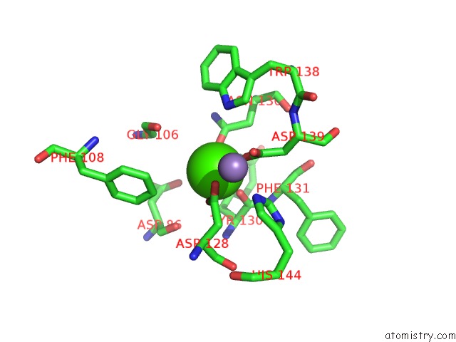
Mono view
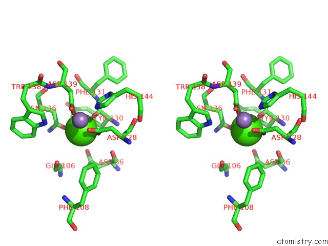
Stereo pair view
Mono view
Stereo pair view
A full contact list of Calcium with other atoms in the Ca binding
site number 1 of Lectin Uea-II Complexed with Chitobiose within 5.0Å range:
|
Calcium binding site 2 out of 2 in 1qos
Go back to
Calcium binding site 2 out
of 2 in the Lectin Uea-II Complexed with Chitobiose
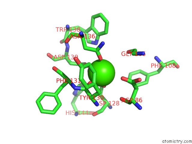
Mono view
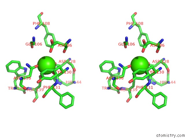
Stereo pair view
Mono view
Stereo pair view
A full contact list of Calcium with other atoms in the Ca binding
site number 2 of Lectin Uea-II Complexed with Chitobiose within 5.0Å range:
|
Reference:
R.Loris,
H.De Greve,
M.-H.Dao-Thi,
J.Messens,
A.Imberty,
L.Wyns.
Structural Basis of Carbohydrate Recognition By Lectin II From Ulex Europaeus, A Protein with A Promiscuous Carbohydrate Binding Site J.Mol.Biol. V. 301 987 2000.
ISSN: ISSN 0022-2836
PubMed: 10966800
DOI: 10.1006/JMBI.2000.4016
Page generated: Tue Jul 8 01:26:55 2025
ISSN: ISSN 0022-2836
PubMed: 10966800
DOI: 10.1006/JMBI.2000.4016
Last articles
Mg in 1VQPMg in 1VQO
Mg in 1W55
Mg in 1W54
Mg in 1W4B
Mg in 1W49
Mg in 1W46
Mg in 1W2Y
Mg in 1VQN
Mg in 1W25