Calcium »
PDB 1rc8-1rpk »
1rir »
Calcium in PDB 1rir: Crystal Structure of Meso-Tetrasulphonatophenylporphyrin in Complex with Peanut Lectin.
Protein crystallography data
The structure of Crystal Structure of Meso-Tetrasulphonatophenylporphyrin in Complex with Peanut Lectin., PDB code: 1rir
was solved by
M.Goel,
K.J.Kaur,
B.G.Maiya,
M.J.Swamy,
D.M.Salunke,
with X-Ray Crystallography technique. A brief refinement statistics is given in the table below:
| Resolution Low / High (Å) | 100.00 / 2.90 |
| Space group | P 32 |
| Cell size a, b, c (Å), α, β, γ (°) | 94.800, 94.800, 144.000, 90.00, 90.00, 120.00 |
| R / Rfree (%) | 23.4 / 28.2 |
Other elements in 1rir:
The structure of Crystal Structure of Meso-Tetrasulphonatophenylporphyrin in Complex with Peanut Lectin. also contains other interesting chemical elements:
| Manganese | (Mn) | 4 atoms |
Calcium Binding Sites:
The binding sites of Calcium atom in the Crystal Structure of Meso-Tetrasulphonatophenylporphyrin in Complex with Peanut Lectin.
(pdb code 1rir). This binding sites where shown within
5.0 Angstroms radius around Calcium atom.
In total 4 binding sites of Calcium where determined in the Crystal Structure of Meso-Tetrasulphonatophenylporphyrin in Complex with Peanut Lectin., PDB code: 1rir:
Jump to Calcium binding site number: 1; 2; 3; 4;
In total 4 binding sites of Calcium where determined in the Crystal Structure of Meso-Tetrasulphonatophenylporphyrin in Complex with Peanut Lectin., PDB code: 1rir:
Jump to Calcium binding site number: 1; 2; 3; 4;
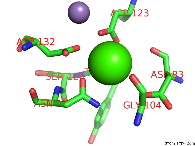
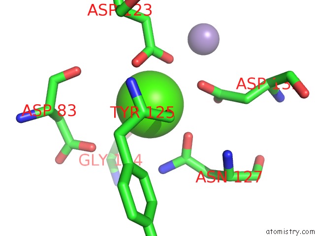
Calcium binding site 1 out of 4 in 1rir
Go back to
Calcium binding site 1 out
of 4 in the Crystal Structure of Meso-Tetrasulphonatophenylporphyrin in Complex with Peanut Lectin.
Mono view
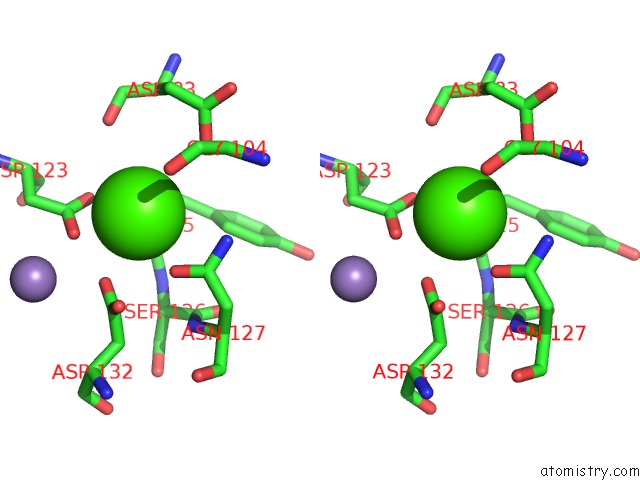
Stereo pair view
Mono view
Stereo pair view
A full contact list of Calcium with other atoms in the Ca binding
site number 1 of Crystal Structure of Meso-Tetrasulphonatophenylporphyrin in Complex with Peanut Lectin. within 5.0Å range:
|
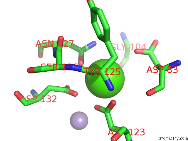
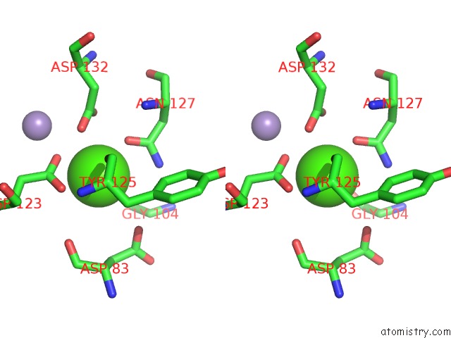
Calcium binding site 2 out of 4 in 1rir
Go back to
Calcium binding site 2 out
of 4 in the Crystal Structure of Meso-Tetrasulphonatophenylporphyrin in Complex with Peanut Lectin.
Mono view
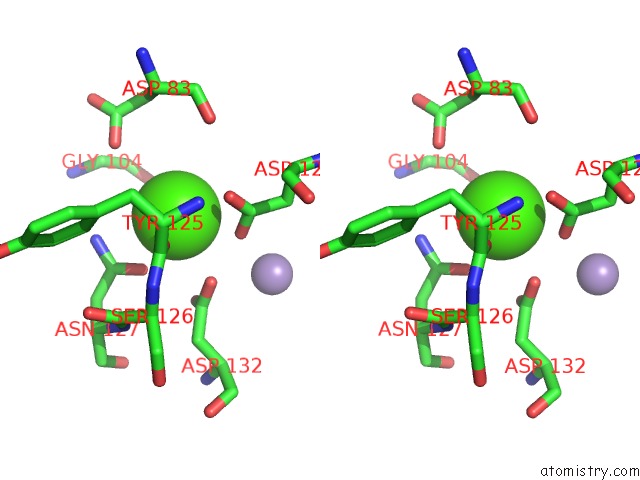
Stereo pair view
Mono view
Stereo pair view
A full contact list of Calcium with other atoms in the Ca binding
site number 2 of Crystal Structure of Meso-Tetrasulphonatophenylporphyrin in Complex with Peanut Lectin. within 5.0Å range:
|
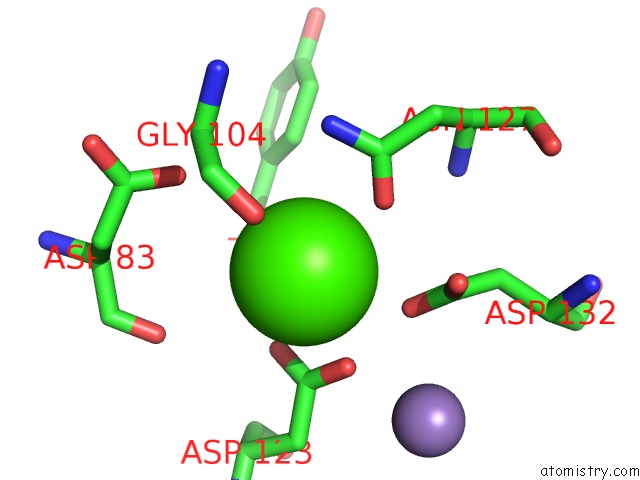
Calcium binding site 3 out of 4 in 1rir
Go back to
Calcium binding site 3 out
of 4 in the Crystal Structure of Meso-Tetrasulphonatophenylporphyrin in Complex with Peanut Lectin.
Mono view
Stereo pair view
Mono view
Stereo pair view
A full contact list of Calcium with other atoms in the Ca binding
site number 3 of Crystal Structure of Meso-Tetrasulphonatophenylporphyrin in Complex with Peanut Lectin. within 5.0Å range:
|
Calcium binding site 4 out of 4 in 1rir
Go back to
Calcium binding site 4 out
of 4 in the Crystal Structure of Meso-Tetrasulphonatophenylporphyrin in Complex with Peanut Lectin.
Mono view
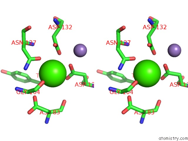
Stereo pair view
Mono view
Stereo pair view
A full contact list of Calcium with other atoms in the Ca binding
site number 4 of Crystal Structure of Meso-Tetrasulphonatophenylporphyrin in Complex with Peanut Lectin. within 5.0Å range:
|
Reference:
M.Goel,
R.S.Damai,
D.K.Sethi,
K.J.Kaur,
B.G.Maiya,
M.J.Swamy,
D.M.Salunke.
Crystal Structures of the Pna-Porphyrin Complex in the Presence and Absence of Lactose: Mapping the Conformational Changes on Lactose Binding, Interacting Surfaces, and Supramolecular Aggregations. Biochemistry V. 44 5588 2005.
ISSN: ISSN 0006-2960
PubMed: 15823017
DOI: 10.1021/BI047377S
Page generated: Tue Jul 8 01:38:13 2025
ISSN: ISSN 0006-2960
PubMed: 15823017
DOI: 10.1021/BI047377S
Last articles
K in 3C33K in 3BO4
K in 3C0J
K in 3BWP
K in 3BOL
K in 3BOF
K in 3BWM
K in 3BEH
K in 3BNS
K in 3BO3