Calcium »
PDB 1rpz-1s26 »
1rx1 »
Calcium in PDB 1rx1: Dihydrofolate Reductase (E.C.1.5.1.3) Complexed with Nicotinamide Adenine Dinucleotide Phosphate (Reduced Form)
Enzymatic activity of Dihydrofolate Reductase (E.C.1.5.1.3) Complexed with Nicotinamide Adenine Dinucleotide Phosphate (Reduced Form)
All present enzymatic activity of Dihydrofolate Reductase (E.C.1.5.1.3) Complexed with Nicotinamide Adenine Dinucleotide Phosphate (Reduced Form):
1.5.1.3;
1.5.1.3;
Protein crystallography data
The structure of Dihydrofolate Reductase (E.C.1.5.1.3) Complexed with Nicotinamide Adenine Dinucleotide Phosphate (Reduced Form), PDB code: 1rx1
was solved by
M.R.Sawaya,
with X-Ray Crystallography technique. A brief refinement statistics is given in the table below:
| Resolution Low / High (Å) | 20.00 / 2.00 |
| Space group | P 21 21 21 |
| Cell size a, b, c (Å), α, β, γ (°) | 34.455, 45.370, 98.701, 90.00, 90.00, 90.00 |
| R / Rfree (%) | 18 / n/a |
Calcium Binding Sites:
The binding sites of Calcium atom in the Dihydrofolate Reductase (E.C.1.5.1.3) Complexed with Nicotinamide Adenine Dinucleotide Phosphate (Reduced Form)
(pdb code 1rx1). This binding sites where shown within
5.0 Angstroms radius around Calcium atom.
In total only one binding site of Calcium was determined in the Dihydrofolate Reductase (E.C.1.5.1.3) Complexed with Nicotinamide Adenine Dinucleotide Phosphate (Reduced Form), PDB code: 1rx1:
In total only one binding site of Calcium was determined in the Dihydrofolate Reductase (E.C.1.5.1.3) Complexed with Nicotinamide Adenine Dinucleotide Phosphate (Reduced Form), PDB code: 1rx1:
Calcium binding site 1 out of 1 in 1rx1
Go back to
Calcium binding site 1 out
of 1 in the Dihydrofolate Reductase (E.C.1.5.1.3) Complexed with Nicotinamide Adenine Dinucleotide Phosphate (Reduced Form)
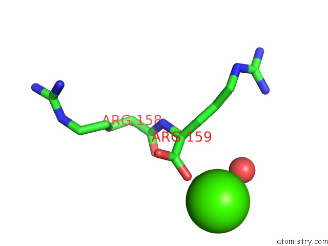
Mono view
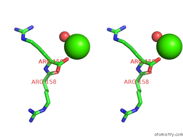
Stereo pair view
Mono view
Stereo pair view
A full contact list of Calcium with other atoms in the Ca binding
site number 1 of Dihydrofolate Reductase (E.C.1.5.1.3) Complexed with Nicotinamide Adenine Dinucleotide Phosphate (Reduced Form) within 5.0Å range:
|
Reference:
M.R.Sawaya,
J.Kraut.
Loop and Subdomain Movements in the Mechanism of Escherichia Coli Dihydrofolate Reductase: Crystallographic Evidence. Biochemistry V. 36 586 1997.
ISSN: ISSN 0006-2960
PubMed: 9012674
DOI: 10.1021/BI962337C
Page generated: Tue Jul 8 01:43:00 2025
ISSN: ISSN 0006-2960
PubMed: 9012674
DOI: 10.1021/BI962337C
Last articles
Mg in 5ZKJMg in 5ZKI
Mg in 5ZK6
Mg in 5ZE9
Mg in 5ZFX
Mg in 5ZCT
Mg in 5ZE6
Mg in 5ZE4
Mg in 5ZDN
Mg in 5ZE0