Calcium »
PDB 1rpz-1s26 »
1s0n »
Calcium in PDB 1s0n: Snapshots of Replication Through An Abasic Lesion: Structural Basis For Base Substitution and Frameshift
Enzymatic activity of Snapshots of Replication Through An Abasic Lesion: Structural Basis For Base Substitution and Frameshift
All present enzymatic activity of Snapshots of Replication Through An Abasic Lesion: Structural Basis For Base Substitution and Frameshift:
2.7.7.7;
2.7.7.7;
Protein crystallography data
The structure of Snapshots of Replication Through An Abasic Lesion: Structural Basis For Base Substitution and Frameshift, PDB code: 1s0n
was solved by
H.Ling,
F.Boudsocq,
R.Woodgate,
W.Yang,
with X-Ray Crystallography technique. A brief refinement statistics is given in the table below:
| Resolution Low / High (Å) | 30.00 / 2.80 |
| Space group | P 21 21 2 |
| Cell size a, b, c (Å), α, β, γ (°) | 99.112, 102.660, 53.176, 90.00, 90.00, 90.00 |
| R / Rfree (%) | 23.8 / 27.5 |
Calcium Binding Sites:
The binding sites of Calcium atom in the Snapshots of Replication Through An Abasic Lesion: Structural Basis For Base Substitution and Frameshift
(pdb code 1s0n). This binding sites where shown within
5.0 Angstroms radius around Calcium atom.
In total 3 binding sites of Calcium where determined in the Snapshots of Replication Through An Abasic Lesion: Structural Basis For Base Substitution and Frameshift, PDB code: 1s0n:
Jump to Calcium binding site number: 1; 2; 3;
In total 3 binding sites of Calcium where determined in the Snapshots of Replication Through An Abasic Lesion: Structural Basis For Base Substitution and Frameshift, PDB code: 1s0n:
Jump to Calcium binding site number: 1; 2; 3;
Calcium binding site 1 out of 3 in 1s0n
Go back to
Calcium binding site 1 out
of 3 in the Snapshots of Replication Through An Abasic Lesion: Structural Basis For Base Substitution and Frameshift

Mono view
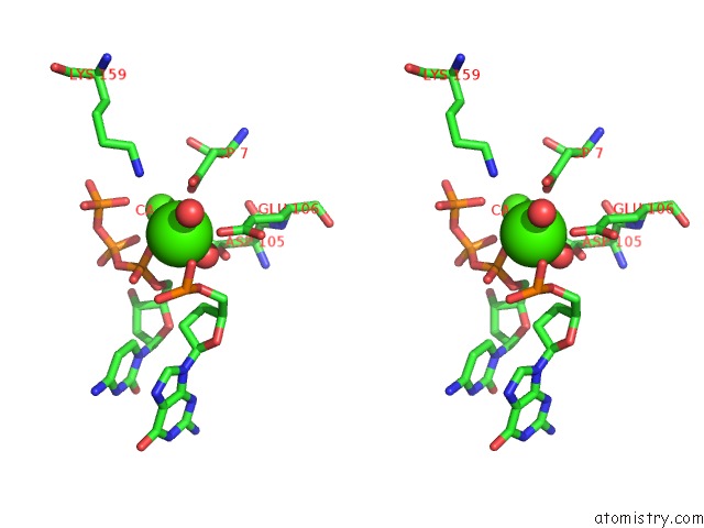
Stereo pair view
Mono view
Stereo pair view
A full contact list of Calcium with other atoms in the Ca binding
site number 1 of Snapshots of Replication Through An Abasic Lesion: Structural Basis For Base Substitution and Frameshift within 5.0Å range:
|
Calcium binding site 2 out of 3 in 1s0n
Go back to
Calcium binding site 2 out
of 3 in the Snapshots of Replication Through An Abasic Lesion: Structural Basis For Base Substitution and Frameshift
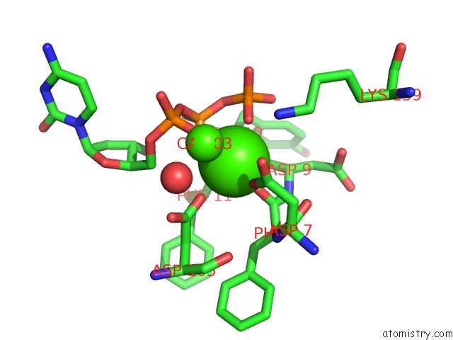
Mono view

Stereo pair view
Mono view
Stereo pair view
A full contact list of Calcium with other atoms in the Ca binding
site number 2 of Snapshots of Replication Through An Abasic Lesion: Structural Basis For Base Substitution and Frameshift within 5.0Å range:
|
Calcium binding site 3 out of 3 in 1s0n
Go back to
Calcium binding site 3 out
of 3 in the Snapshots of Replication Through An Abasic Lesion: Structural Basis For Base Substitution and Frameshift
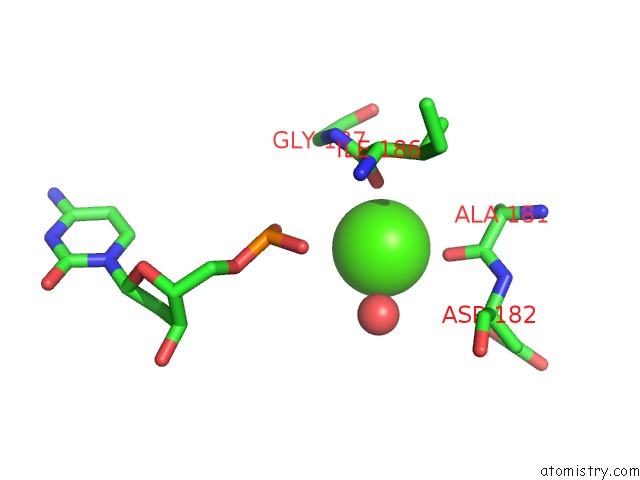
Mono view

Stereo pair view
Mono view
Stereo pair view
A full contact list of Calcium with other atoms in the Ca binding
site number 3 of Snapshots of Replication Through An Abasic Lesion: Structural Basis For Base Substitution and Frameshift within 5.0Å range:
|
Reference:
H.Ling,
F.Boudsocq,
R.Woodgate,
W.Yang.
Snapshots of Replication Through An Abasic Lesion; Structural Basis For Base Substitutions and Frameshifts. Mol.Cell V. 13 751 2004.
ISSN: ISSN 1097-2765
PubMed: 15023344
DOI: 10.1016/S1097-2765(04)00101-7
Page generated: Tue Jul 8 01:45:53 2025
ISSN: ISSN 1097-2765
PubMed: 15023344
DOI: 10.1016/S1097-2765(04)00101-7
Last articles
Mg in 6DLJMg in 6DJQ
Mg in 6DJO
Mg in 6DJN
Mg in 6DJM
Mg in 6DGI
Mg in 6DFF
Mg in 6DHT
Mg in 6DI7
Mg in 6DEX