Calcium »
PDB 1s2n-1scn »
1scm »
Calcium in PDB 1scm: Structure of the Regulatory Domain of Scallop Myosin at 2.8 Angstroms Resolution
Protein crystallography data
The structure of Structure of the Regulatory Domain of Scallop Myosin at 2.8 Angstroms Resolution, PDB code: 1scm
was solved by
C.Cohen,
X.Xie,
with X-Ray Crystallography technique. A brief refinement statistics is given in the table below:
| Resolution Low / High (Å) | 10.00 / 2.80 |
| Space group | P 1 21 1 |
| Cell size a, b, c (Å), α, β, γ (°) | 52.500, 87.000, 55.500, 90.00, 114.50, 90.00 |
| R / Rfree (%) | 20.1 / n/a |
Calcium Binding Sites:
The binding sites of Calcium atom in the Structure of the Regulatory Domain of Scallop Myosin at 2.8 Angstroms Resolution
(pdb code 1scm). This binding sites where shown within
5.0 Angstroms radius around Calcium atom.
In total 2 binding sites of Calcium where determined in the Structure of the Regulatory Domain of Scallop Myosin at 2.8 Angstroms Resolution, PDB code: 1scm:
Jump to Calcium binding site number: 1; 2;
In total 2 binding sites of Calcium where determined in the Structure of the Regulatory Domain of Scallop Myosin at 2.8 Angstroms Resolution, PDB code: 1scm:
Jump to Calcium binding site number: 1; 2;
Calcium binding site 1 out of 2 in 1scm
Go back to
Calcium binding site 1 out
of 2 in the Structure of the Regulatory Domain of Scallop Myosin at 2.8 Angstroms Resolution
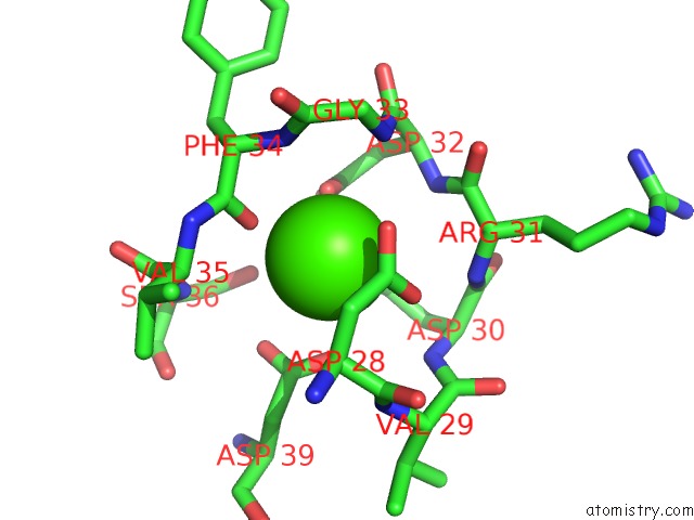
Mono view
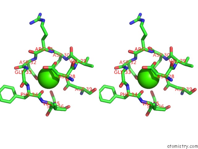
Stereo pair view
Mono view
Stereo pair view
A full contact list of Calcium with other atoms in the Ca binding
site number 1 of Structure of the Regulatory Domain of Scallop Myosin at 2.8 Angstroms Resolution within 5.0Å range:
|
Calcium binding site 2 out of 2 in 1scm
Go back to
Calcium binding site 2 out
of 2 in the Structure of the Regulatory Domain of Scallop Myosin at 2.8 Angstroms Resolution
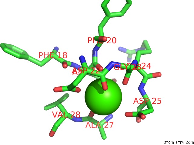
Mono view
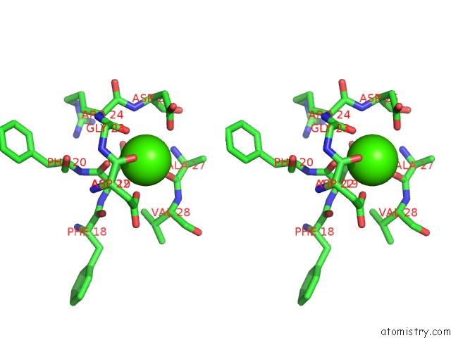
Stereo pair view
Mono view
Stereo pair view
A full contact list of Calcium with other atoms in the Ca binding
site number 2 of Structure of the Regulatory Domain of Scallop Myosin at 2.8 Angstroms Resolution within 5.0Å range:
|
Reference:
X.Xie,
D.H.Harrison,
I.Schlichting,
R.M.Sweet,
V.N.Kalabokis,
A.G.Szent-Gyorgyi,
C.Cohen.
Structure of the Regulatory Domain of Scallop Myosin at 2.8 A Resolution. Nature V. 368 306 1994.
ISSN: ISSN 0028-0836
PubMed: 8127365
DOI: 10.1038/368306A0
Page generated: Tue Jul 8 01:54:23 2025
ISSN: ISSN 0028-0836
PubMed: 8127365
DOI: 10.1038/368306A0
Last articles
Mg in 2PANMg in 2PI4
Mg in 2PGO
Mg in 2PGN
Mg in 2PG2
Mg in 2PFQ
Mg in 2PA4
Mg in 2PFP
Mg in 2PFO
Mg in 2PDA