Calcium »
PDB 1scr-1spj »
1sln »
Calcium in PDB 1sln: Crystal Structure of the Catalytic Domain of Human Fibroblast Stromelysin-1 Inhibited with the N-Carboxy-Alkyl Inhibitor L-702,842
Enzymatic activity of Crystal Structure of the Catalytic Domain of Human Fibroblast Stromelysin-1 Inhibited with the N-Carboxy-Alkyl Inhibitor L-702,842
All present enzymatic activity of Crystal Structure of the Catalytic Domain of Human Fibroblast Stromelysin-1 Inhibited with the N-Carboxy-Alkyl Inhibitor L-702,842:
3.4.24.17;
3.4.24.17;
Protein crystallography data
The structure of Crystal Structure of the Catalytic Domain of Human Fibroblast Stromelysin-1 Inhibited with the N-Carboxy-Alkyl Inhibitor L-702,842, PDB code: 1sln
was solved by
J.W.Becker,
with X-Ray Crystallography technique. A brief refinement statistics is given in the table below:
| Resolution Low / High (Å) | 8.00 / 2.27 |
| Space group | P 31 2 1 |
| Cell size a, b, c (Å), α, β, γ (°) | 47.230, 47.230, 150.850, 90.00, 90.00, 120.00 |
| R / Rfree (%) | 22.6 / 29.9 |
Other elements in 1sln:
The structure of Crystal Structure of the Catalytic Domain of Human Fibroblast Stromelysin-1 Inhibited with the N-Carboxy-Alkyl Inhibitor L-702,842 also contains other interesting chemical elements:
| Zinc | (Zn) | 2 atoms |
Calcium Binding Sites:
The binding sites of Calcium atom in the Crystal Structure of the Catalytic Domain of Human Fibroblast Stromelysin-1 Inhibited with the N-Carboxy-Alkyl Inhibitor L-702,842
(pdb code 1sln). This binding sites where shown within
5.0 Angstroms radius around Calcium atom.
In total 3 binding sites of Calcium where determined in the Crystal Structure of the Catalytic Domain of Human Fibroblast Stromelysin-1 Inhibited with the N-Carboxy-Alkyl Inhibitor L-702,842, PDB code: 1sln:
Jump to Calcium binding site number: 1; 2; 3;
In total 3 binding sites of Calcium where determined in the Crystal Structure of the Catalytic Domain of Human Fibroblast Stromelysin-1 Inhibited with the N-Carboxy-Alkyl Inhibitor L-702,842, PDB code: 1sln:
Jump to Calcium binding site number: 1; 2; 3;
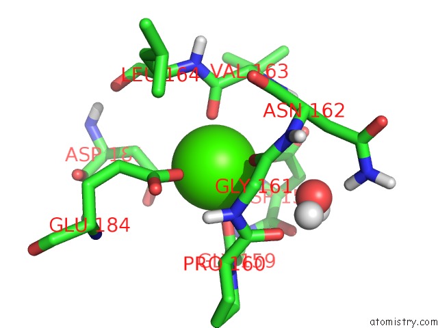
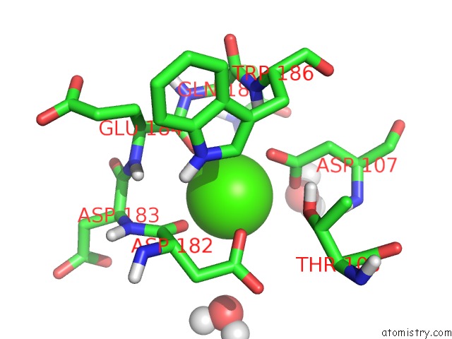
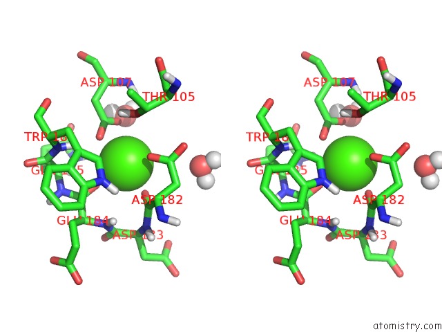
Calcium binding site 1 out of 3 in 1sln
Go back to
Calcium binding site 1 out
of 3 in the Crystal Structure of the Catalytic Domain of Human Fibroblast Stromelysin-1 Inhibited with the N-Carboxy-Alkyl Inhibitor L-702,842
Mono view
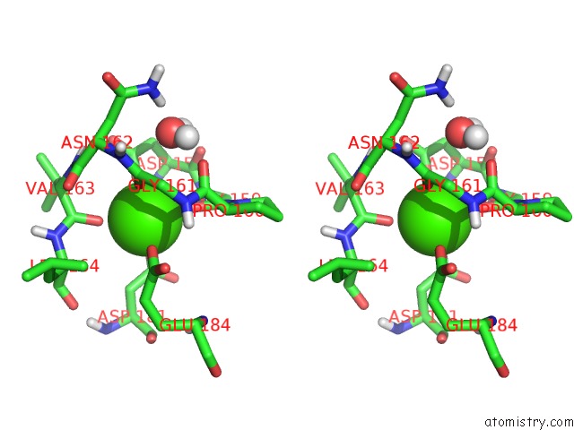
Stereo pair view
Mono view
Stereo pair view
A full contact list of Calcium with other atoms in the Ca binding
site number 1 of Crystal Structure of the Catalytic Domain of Human Fibroblast Stromelysin-1 Inhibited with the N-Carboxy-Alkyl Inhibitor L-702,842 within 5.0Å range:
|
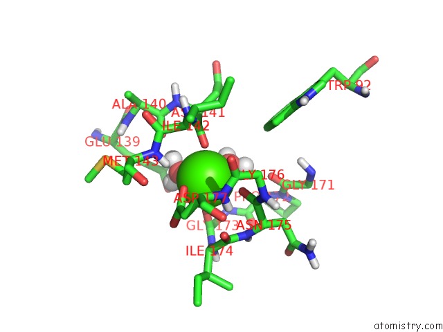
Calcium binding site 2 out of 3 in 1sln
Go back to
Calcium binding site 2 out
of 3 in the Crystal Structure of the Catalytic Domain of Human Fibroblast Stromelysin-1 Inhibited with the N-Carboxy-Alkyl Inhibitor L-702,842
Mono view
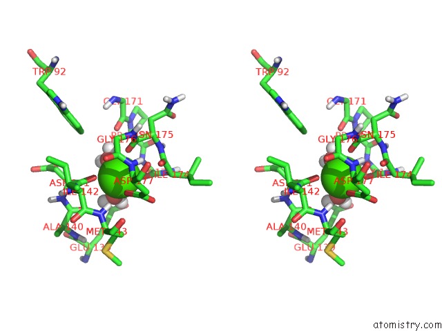
Stereo pair view
Mono view
Stereo pair view
A full contact list of Calcium with other atoms in the Ca binding
site number 2 of Crystal Structure of the Catalytic Domain of Human Fibroblast Stromelysin-1 Inhibited with the N-Carboxy-Alkyl Inhibitor L-702,842 within 5.0Å range:
|
Calcium binding site 3 out of 3 in 1sln
Go back to
Calcium binding site 3 out
of 3 in the Crystal Structure of the Catalytic Domain of Human Fibroblast Stromelysin-1 Inhibited with the N-Carboxy-Alkyl Inhibitor L-702,842
Mono view
Stereo pair view
Mono view
Stereo pair view
A full contact list of Calcium with other atoms in the Ca binding
site number 3 of Crystal Structure of the Catalytic Domain of Human Fibroblast Stromelysin-1 Inhibited with the N-Carboxy-Alkyl Inhibitor L-702,842 within 5.0Å range:
|
Reference:
J.W.Becker,
A.I.Marcy,
L.L.Rokosz,
M.G.Axel,
J.J.Burbaum,
P.M.Fitzgerald,
P.M.Cameron,
C.K.Esser,
W.K.Hagmann,
J.D.Hermes,
J.P.Springer.
Stromelysin-1: Three-Dimensional Structure of the Inhibited Catalytic Domain and of the C-Truncated Proenzyme. Protein Sci. V. 4 1966 1995.
ISSN: ISSN 0961-8368
PubMed: 8535233
Page generated: Tue Jul 8 02:00:25 2025
ISSN: ISSN 0961-8368
PubMed: 8535233
Last articles
K in 7OV7K in 7OTJ
K in 7OTB
K in 7OPH
K in 7OUP
K in 7OUE
K in 7OQT
K in 7OQ1
K in 7OT4
K in 7OOU