Calcium »
PDB 1spu-1t5s »
1svy »
Calcium in PDB 1svy: Severin Domain 2, 1.75 Angstrom Crystal Structure
Protein crystallography data
The structure of Severin Domain 2, 1.75 Angstrom Crystal Structure, PDB code: 1svy
was solved by
Y.A.Puius,
E.V.Fedorov,
L.Eichinger,
M.Sullivan,
M.Schleicher,
S.C.Almo,
with X-Ray Crystallography technique. A brief refinement statistics is given in the table below:
| Resolution Low / High (Å) | 20.00 / 1.75 |
| Space group | P 21 21 21 |
| Cell size a, b, c (Å), α, β, γ (°) | 33.352, 49.218, 62.790, 90.00, 90.00, 90.00 |
| R / Rfree (%) | 18.4 / 24.6 |
Other elements in 1svy:
The structure of Severin Domain 2, 1.75 Angstrom Crystal Structure also contains other interesting chemical elements:
| Sodium | (Na) | 1 atom |
Calcium Binding Sites:
The binding sites of Calcium atom in the Severin Domain 2, 1.75 Angstrom Crystal Structure
(pdb code 1svy). This binding sites where shown within
5.0 Angstroms radius around Calcium atom.
In total only one binding site of Calcium was determined in the Severin Domain 2, 1.75 Angstrom Crystal Structure, PDB code: 1svy:
In total only one binding site of Calcium was determined in the Severin Domain 2, 1.75 Angstrom Crystal Structure, PDB code: 1svy:
Calcium binding site 1 out of 1 in 1svy
Go back to
Calcium binding site 1 out
of 1 in the Severin Domain 2, 1.75 Angstrom Crystal Structure
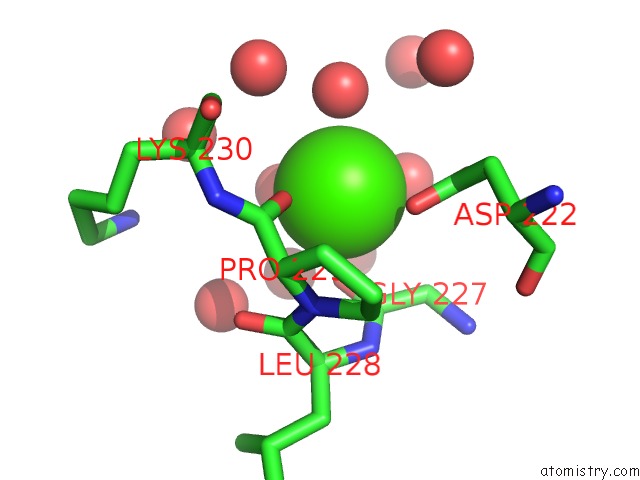
Mono view
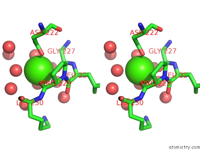
Stereo pair view
Mono view
Stereo pair view
A full contact list of Calcium with other atoms in the Ca binding
site number 1 of Severin Domain 2, 1.75 Angstrom Crystal Structure within 5.0Å range:
|
Reference:
Y.A.Puius,
E.V.Fedorov,
L.Eichinger,
M.Schleicher,
S.C.Almo.
Mapping the Functional Surface of Domain 2 in the Gelsolin Superfamily. Biochemistry V. 39 5322 2000.
ISSN: ISSN 0006-2960
PubMed: 10820002
DOI: 10.1021/BI992364D
Page generated: Tue Jul 8 02:07:18 2025
ISSN: ISSN 0006-2960
PubMed: 10820002
DOI: 10.1021/BI992364D
Last articles
Na in 3MUANa in 3MUI
Na in 3MUX
Na in 3MU8
Na in 3MU5
Na in 3MU4
Na in 3MU1
Na in 3MSG
Na in 3MU0
Na in 3MR1