Calcium »
PDB 1t5t-1tkh »
1tk2 »
Calcium in PDB 1tk2: Crystal Structure of the Complex Formed Between Alkaline Proteinase Savinase and Gramicidin S at 1.5A Resolution
Enzymatic activity of Crystal Structure of the Complex Formed Between Alkaline Proteinase Savinase and Gramicidin S at 1.5A Resolution
All present enzymatic activity of Crystal Structure of the Complex Formed Between Alkaline Proteinase Savinase and Gramicidin S at 1.5A Resolution:
3.4.21.62;
3.4.21.62;
Protein crystallography data
The structure of Crystal Structure of the Complex Formed Between Alkaline Proteinase Savinase and Gramicidin S at 1.5A Resolution, PDB code: 1tk2
was solved by
V.S.Bhatt,
P.Kaur,
S.Klupsch,
C.Betzel,
S.Brenner,
T.P.Singh,
with X-Ray Crystallography technique. A brief refinement statistics is given in the table below:
| Resolution Low / High (Å) | 27.32 / 1.54 |
| Space group | P 21 21 21 |
| Cell size a, b, c (Å), α, β, γ (°) | 76.250, 73.340, 40.890, 90.00, 90.00, 90.00 |
| R / Rfree (%) | 16.9 / 18.9 |
Calcium Binding Sites:
The binding sites of Calcium atom in the Crystal Structure of the Complex Formed Between Alkaline Proteinase Savinase and Gramicidin S at 1.5A Resolution
(pdb code 1tk2). This binding sites where shown within
5.0 Angstroms radius around Calcium atom.
In total 2 binding sites of Calcium where determined in the Crystal Structure of the Complex Formed Between Alkaline Proteinase Savinase and Gramicidin S at 1.5A Resolution, PDB code: 1tk2:
Jump to Calcium binding site number: 1; 2;
In total 2 binding sites of Calcium where determined in the Crystal Structure of the Complex Formed Between Alkaline Proteinase Savinase and Gramicidin S at 1.5A Resolution, PDB code: 1tk2:
Jump to Calcium binding site number: 1; 2;
Calcium binding site 1 out of 2 in 1tk2
Go back to
Calcium binding site 1 out
of 2 in the Crystal Structure of the Complex Formed Between Alkaline Proteinase Savinase and Gramicidin S at 1.5A Resolution
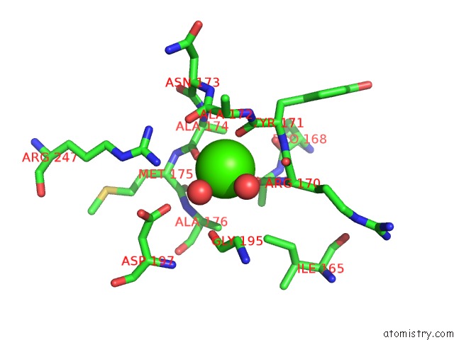
Mono view
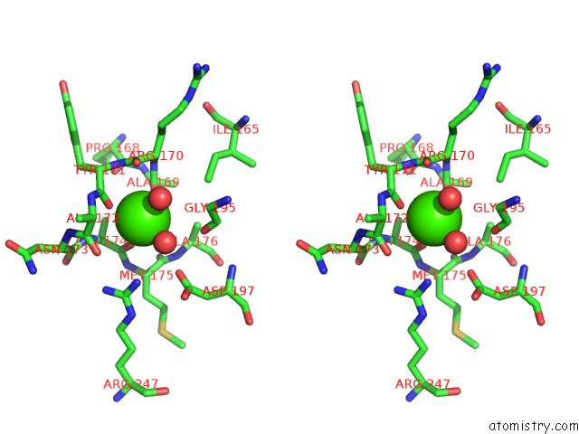
Stereo pair view
Mono view
Stereo pair view
A full contact list of Calcium with other atoms in the Ca binding
site number 1 of Crystal Structure of the Complex Formed Between Alkaline Proteinase Savinase and Gramicidin S at 1.5A Resolution within 5.0Å range:
|
Calcium binding site 2 out of 2 in 1tk2
Go back to
Calcium binding site 2 out
of 2 in the Crystal Structure of the Complex Formed Between Alkaline Proteinase Savinase and Gramicidin S at 1.5A Resolution
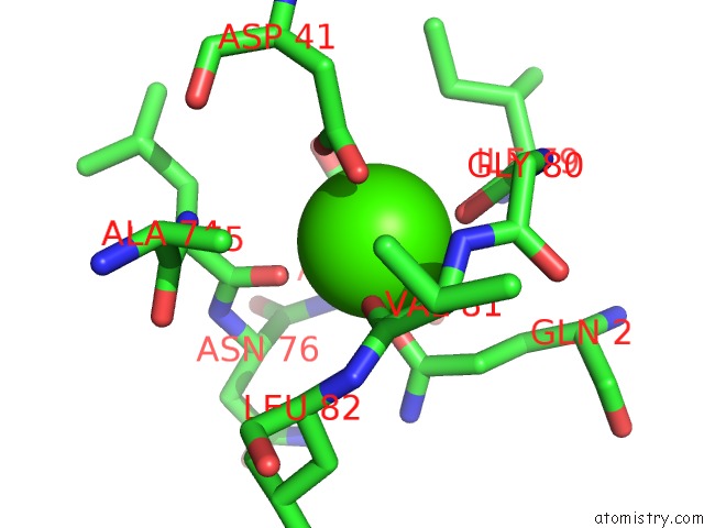
Mono view
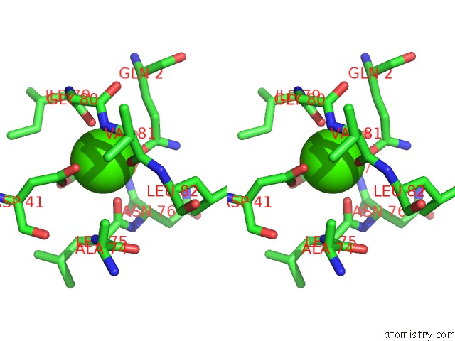
Stereo pair view
Mono view
Stereo pair view
A full contact list of Calcium with other atoms in the Ca binding
site number 2 of Crystal Structure of the Complex Formed Between Alkaline Proteinase Savinase and Gramicidin S at 1.5A Resolution within 5.0Å range:
|
Reference:
V.S.Bhatt,
P.Kaur,
S.Klupsch,
C.Betzel,
S.Brenner,
T.P.Singh.
Crystal Structure of the Complex Formed Between Alkaline Proteinase Savinase and Gramicidin S at 1.5A Resolution. To Be Published.
Page generated: Tue Jul 8 02:16:02 2025
Last articles
Mg in 1Z5CMg in 1Z5A
Mg in 1Z4Q
Mg in 1Z59
Mg in 1Z4O
Mg in 1Z4P
Mg in 1Z4N
Mg in 1Z4L
Mg in 1Z4M
Mg in 1Z4J