Calcium »
PDB 1tu5-1ujc »
1u5q »
Calcium in PDB 1u5q: Crystal Structure of the TAO2 Kinase Domain: Activation and Specifity of A STE20P MAP3K
Protein crystallography data
The structure of Crystal Structure of the TAO2 Kinase Domain: Activation and Specifity of A STE20P MAP3K, PDB code: 1u5q
was solved by
T.Zhou,
M.Raman,
Y.Gao,
S.Earnest,
Z.Chen,
M.Machius,
M.H.Cobb,
E.J.Goldsmith,
with X-Ray Crystallography technique. A brief refinement statistics is given in the table below:
| Resolution Low / High (Å) | 50.00 / 2.10 |
| Space group | P 65 2 2 |
| Cell size a, b, c (Å), α, β, γ (°) | 186.220, 186.220, 94.506, 90.00, 90.00, 120.00 |
| R / Rfree (%) | 22.3 / 26.9 |
Calcium Binding Sites:
The binding sites of Calcium atom in the Crystal Structure of the TAO2 Kinase Domain: Activation and Specifity of A STE20P MAP3K
(pdb code 1u5q). This binding sites where shown within
5.0 Angstroms radius around Calcium atom.
In total 2 binding sites of Calcium where determined in the Crystal Structure of the TAO2 Kinase Domain: Activation and Specifity of A STE20P MAP3K, PDB code: 1u5q:
Jump to Calcium binding site number: 1; 2;
In total 2 binding sites of Calcium where determined in the Crystal Structure of the TAO2 Kinase Domain: Activation and Specifity of A STE20P MAP3K, PDB code: 1u5q:
Jump to Calcium binding site number: 1; 2;
Calcium binding site 1 out of 2 in 1u5q
Go back to
Calcium binding site 1 out
of 2 in the Crystal Structure of the TAO2 Kinase Domain: Activation and Specifity of A STE20P MAP3K
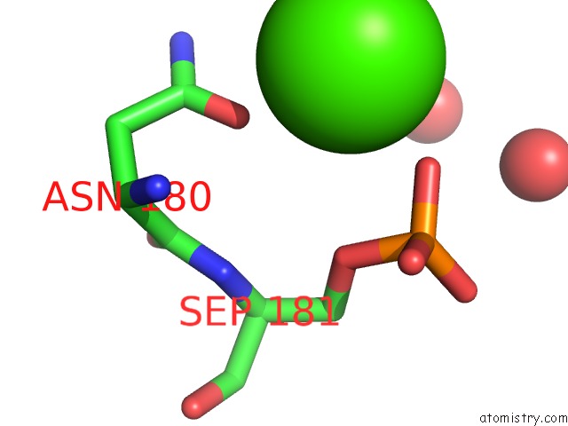
Mono view
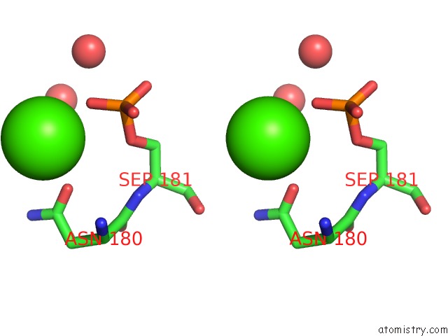
Stereo pair view
Mono view
Stereo pair view
A full contact list of Calcium with other atoms in the Ca binding
site number 1 of Crystal Structure of the TAO2 Kinase Domain: Activation and Specifity of A STE20P MAP3K within 5.0Å range:
|
Calcium binding site 2 out of 2 in 1u5q
Go back to
Calcium binding site 2 out
of 2 in the Crystal Structure of the TAO2 Kinase Domain: Activation and Specifity of A STE20P MAP3K
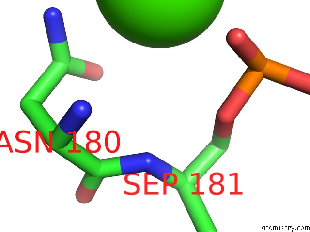
Mono view
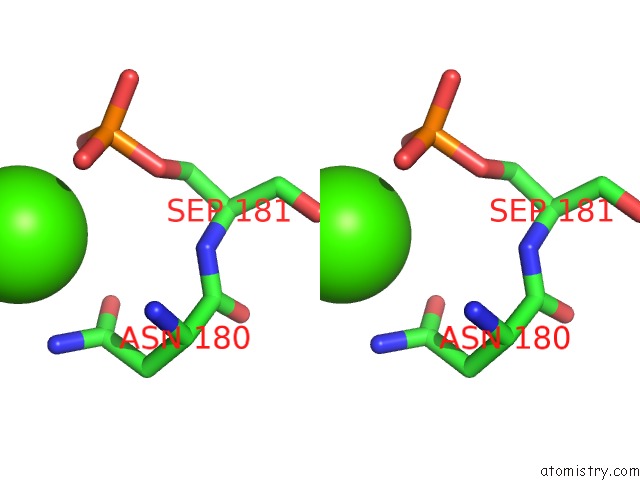
Stereo pair view
Mono view
Stereo pair view
A full contact list of Calcium with other atoms in the Ca binding
site number 2 of Crystal Structure of the TAO2 Kinase Domain: Activation and Specifity of A STE20P MAP3K within 5.0Å range:
|
Reference:
T.Zhou,
M.Raman,
Y.Gao,
S.Earnest,
Z.Chen,
M.Machius,
M.H.Cobb,
E.J.Goldsmith.
Crystal Structure of the TAO2 Kinase Domain; Activation and Specificity of A STE20P MAP3K. Structure V. 12 1891 2004.
ISSN: ISSN 0969-2126
PubMed: 15458637
DOI: 10.1016/J.STR.2004.07.021
Page generated: Tue Jul 8 02:26:21 2025
ISSN: ISSN 0969-2126
PubMed: 15458637
DOI: 10.1016/J.STR.2004.07.021
Last articles
I in 7DCZI in 7D36
I in 7D2V
I in 7D5A
I in 7D2X
I in 7CX9
I in 7CP7
I in 7CRF
I in 7CP6
I in 7BYD