Calcium »
PDB 1tu5-1ujc »
1ua7 »
Calcium in PDB 1ua7: Crystal Structure Analysis of Alpha-Amylase From Bacillus Subtilis Complexed with Acarbose
Enzymatic activity of Crystal Structure Analysis of Alpha-Amylase From Bacillus Subtilis Complexed with Acarbose
All present enzymatic activity of Crystal Structure Analysis of Alpha-Amylase From Bacillus Subtilis Complexed with Acarbose:
3.2.1.1;
3.2.1.1;
Protein crystallography data
The structure of Crystal Structure Analysis of Alpha-Amylase From Bacillus Subtilis Complexed with Acarbose, PDB code: 1ua7
was solved by
M.Kagawa,
Z.Fujimoto,
M.Momma,
K.Takase,
H.Mizuno,
with X-Ray Crystallography technique. A brief refinement statistics is given in the table below:
| Resolution Low / High (Å) | 19.97 / 2.21 |
| Space group | P 21 21 21 |
| Cell size a, b, c (Å), α, β, γ (°) | 70.320, 74.204, 115.724, 90.00, 90.00, 90.00 |
| R / Rfree (%) | 20.8 / 26.5 |
Calcium Binding Sites:
The binding sites of Calcium atom in the Crystal Structure Analysis of Alpha-Amylase From Bacillus Subtilis Complexed with Acarbose
(pdb code 1ua7). This binding sites where shown within
5.0 Angstroms radius around Calcium atom.
In total 3 binding sites of Calcium where determined in the Crystal Structure Analysis of Alpha-Amylase From Bacillus Subtilis Complexed with Acarbose, PDB code: 1ua7:
Jump to Calcium binding site number: 1; 2; 3;
In total 3 binding sites of Calcium where determined in the Crystal Structure Analysis of Alpha-Amylase From Bacillus Subtilis Complexed with Acarbose, PDB code: 1ua7:
Jump to Calcium binding site number: 1; 2; 3;
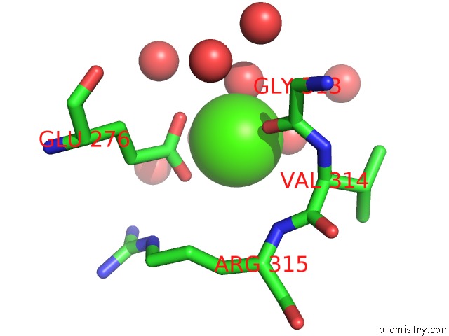
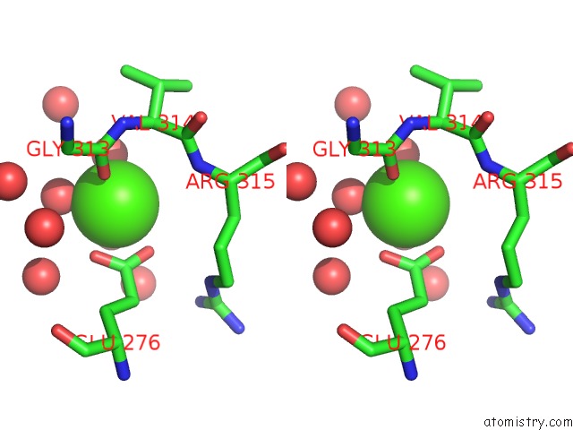
Calcium binding site 1 out of 3 in 1ua7
Go back to
Calcium binding site 1 out
of 3 in the Crystal Structure Analysis of Alpha-Amylase From Bacillus Subtilis Complexed with Acarbose
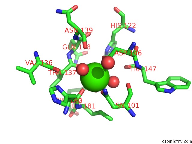
Mono view
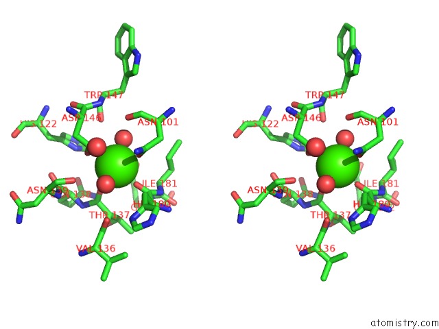
Stereo pair view
Mono view
Stereo pair view
A full contact list of Calcium with other atoms in the Ca binding
site number 1 of Crystal Structure Analysis of Alpha-Amylase From Bacillus Subtilis Complexed with Acarbose within 5.0Å range:
|
Calcium binding site 2 out of 3 in 1ua7
Go back to
Calcium binding site 2 out
of 3 in the Crystal Structure Analysis of Alpha-Amylase From Bacillus Subtilis Complexed with Acarbose
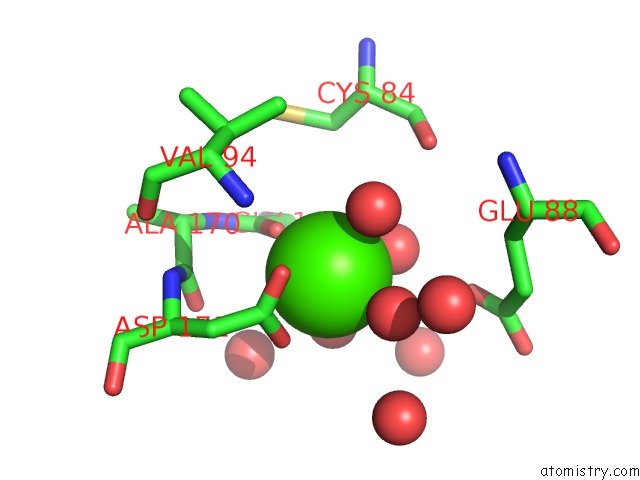
Mono view
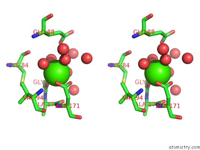
Stereo pair view
Mono view
Stereo pair view
A full contact list of Calcium with other atoms in the Ca binding
site number 2 of Crystal Structure Analysis of Alpha-Amylase From Bacillus Subtilis Complexed with Acarbose within 5.0Å range:
|
Calcium binding site 3 out of 3 in 1ua7
Go back to
Calcium binding site 3 out
of 3 in the Crystal Structure Analysis of Alpha-Amylase From Bacillus Subtilis Complexed with Acarbose
Mono view
Stereo pair view
Mono view
Stereo pair view
A full contact list of Calcium with other atoms in the Ca binding
site number 3 of Crystal Structure Analysis of Alpha-Amylase From Bacillus Subtilis Complexed with Acarbose within 5.0Å range:
|
Reference:
M.Kagawa,
Z.Fujimoto,
M.Momma,
K.Takase,
H.Mizuno.
Crystal Structure of Bacillus Subtilis Alpha-Amylase in Complex with Acarbose J.Bacteriol. V. 185 6981 2003.
ISSN: ISSN 0021-9193
PubMed: 14617662
DOI: 10.1128/JB.185.23.6981-6984.2003
Page generated: Tue Jul 8 02:29:00 2025
ISSN: ISSN 0021-9193
PubMed: 14617662
DOI: 10.1128/JB.185.23.6981-6984.2003
Last articles
K in 1GA9K in 1FPI
K in 1G75
K in 1F3X
K in 1FT7
K in 1FQE
K in 1F3W
K in 1FP7
K in 1FL1
K in 1FFY