Calcium »
PDB 1tu5-1ujc »
1ujb »
Calcium in PDB 1ujb: Structure of the Protein Histidine Phosphatase Sixa
Protein crystallography data
The structure of Structure of the Protein Histidine Phosphatase Sixa, PDB code: 1ujb
was solved by
K.Hamada,
M.Kato,
T.Shimizu,
K.Ihara,
T.Mizuno,
T.Hakoshima,
with X-Ray Crystallography technique. A brief refinement statistics is given in the table below:
| Resolution Low / High (Å) | 41.98 / 2.06 |
| Space group | P 21 21 21 |
| Cell size a, b, c (Å), α, β, γ (°) | 39.260, 48.620, 83.180, 90.00, 90.00, 90.00 |
| R / Rfree (%) | 17.3 / 22.9 |
Calcium Binding Sites:
The binding sites of Calcium atom in the Structure of the Protein Histidine Phosphatase Sixa
(pdb code 1ujb). This binding sites where shown within
5.0 Angstroms radius around Calcium atom.
In total only one binding site of Calcium was determined in the Structure of the Protein Histidine Phosphatase Sixa, PDB code: 1ujb:
In total only one binding site of Calcium was determined in the Structure of the Protein Histidine Phosphatase Sixa, PDB code: 1ujb:
Calcium binding site 1 out of 1 in 1ujb
Go back to
Calcium binding site 1 out
of 1 in the Structure of the Protein Histidine Phosphatase Sixa
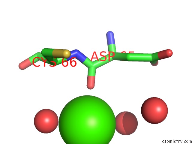
Mono view
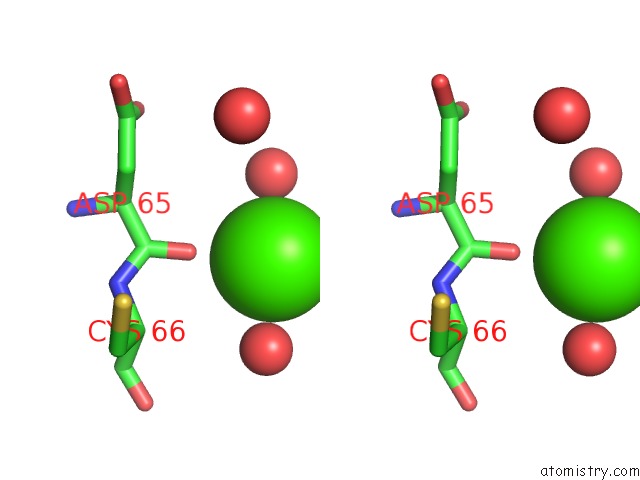
Stereo pair view
Mono view
Stereo pair view
A full contact list of Calcium with other atoms in the Ca binding
site number 1 of Structure of the Protein Histidine Phosphatase Sixa within 5.0Å range:
|
Reference:
K.Hamada,
M.Kato,
T.Shimizu,
K.Ihara,
T.Mizuno,
T.Hakoshima.
Crystal Structure of the Protein Histidine Phosphatase Sixa in the Multistep His-Asp Phosphorelay. Genes Cells V. 10 1 2005.
ISSN: ISSN 1356-9597
PubMed: 15670209
DOI: 10.1111/J.1365-2443.2005.00817.X
Page generated: Tue Jul 8 02:34:43 2025
ISSN: ISSN 1356-9597
PubMed: 15670209
DOI: 10.1111/J.1365-2443.2005.00817.X
Last articles
Mg in 5J34Mg in 5J3T
Mg in 5J3P
Mg in 5J2U
Mg in 5J32
Mg in 5J2T
Mg in 5J2P
Mg in 5J2Q
Mg in 5J02
Mg in 5J01