Calcium »
PDB 1ukg-1ux6 »
1uoc »
Calcium in PDB 1uoc: X-Ray Structure of the Rnase Domain of the Yeast POP2 Protein
Protein crystallography data
The structure of X-Ray Structure of the Rnase Domain of the Yeast POP2 Protein, PDB code: 1uoc
was solved by
S.Thore,
F.Mauxion,
B.Seraphin,
D.Suck,
with X-Ray Crystallography technique. A brief refinement statistics is given in the table below:
| Resolution Low / High (Å) | 9.97 / 2.3 |
| Space group | P 21 21 21 |
| Cell size a, b, c (Å), α, β, γ (°) | 78.582, 79.440, 101.324, 90.00, 90.00, 90.00 |
| R / Rfree (%) | 23.9 / 26.5 |
Other elements in 1uoc:
The structure of X-Ray Structure of the Rnase Domain of the Yeast POP2 Protein also contains other interesting chemical elements:
| Xenon | (Xe) | 2 atoms |
Calcium Binding Sites:
The binding sites of Calcium atom in the X-Ray Structure of the Rnase Domain of the Yeast POP2 Protein
(pdb code 1uoc). This binding sites where shown within
5.0 Angstroms radius around Calcium atom.
In total 2 binding sites of Calcium where determined in the X-Ray Structure of the Rnase Domain of the Yeast POP2 Protein, PDB code: 1uoc:
Jump to Calcium binding site number: 1; 2;
In total 2 binding sites of Calcium where determined in the X-Ray Structure of the Rnase Domain of the Yeast POP2 Protein, PDB code: 1uoc:
Jump to Calcium binding site number: 1; 2;
Calcium binding site 1 out of 2 in 1uoc
Go back to
Calcium binding site 1 out
of 2 in the X-Ray Structure of the Rnase Domain of the Yeast POP2 Protein
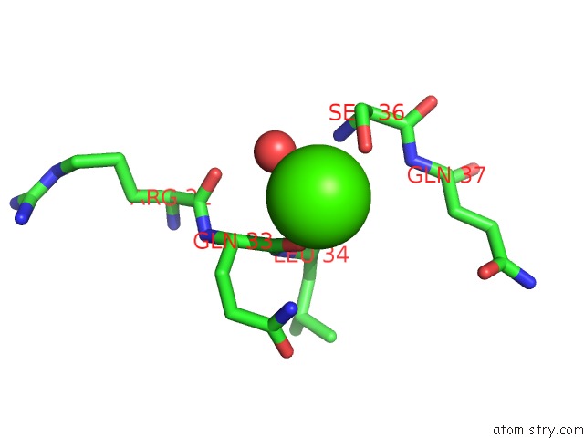
Mono view
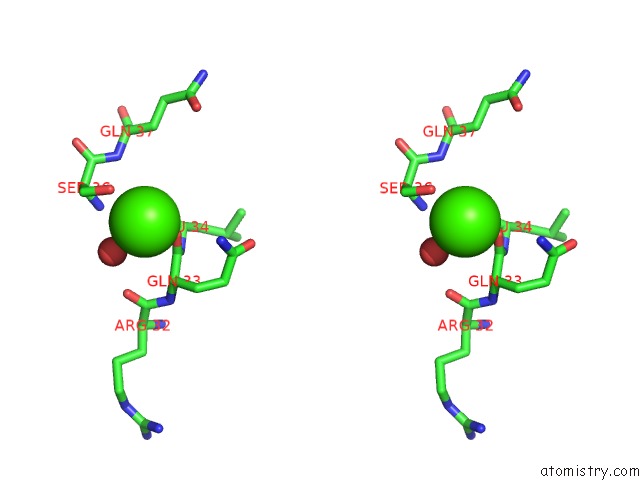
Stereo pair view
Mono view
Stereo pair view
A full contact list of Calcium with other atoms in the Ca binding
site number 1 of X-Ray Structure of the Rnase Domain of the Yeast POP2 Protein within 5.0Å range:
|
Calcium binding site 2 out of 2 in 1uoc
Go back to
Calcium binding site 2 out
of 2 in the X-Ray Structure of the Rnase Domain of the Yeast POP2 Protein
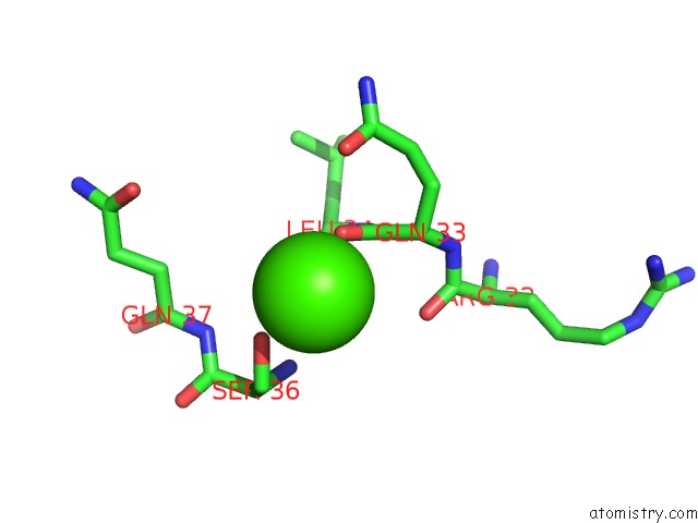
Mono view
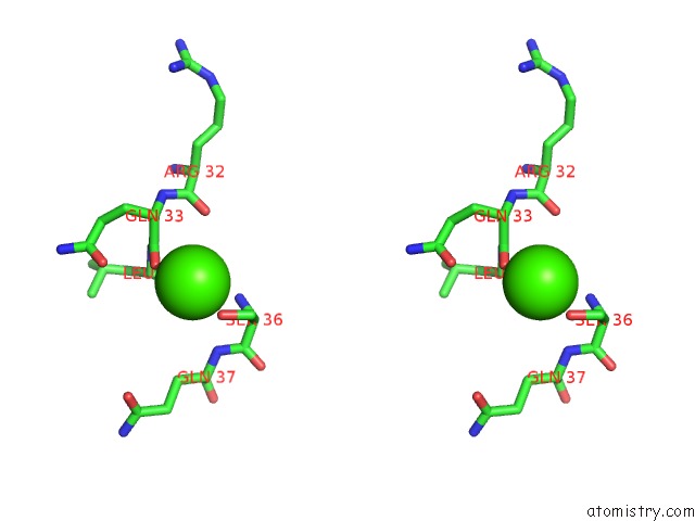
Stereo pair view
Mono view
Stereo pair view
A full contact list of Calcium with other atoms in the Ca binding
site number 2 of X-Ray Structure of the Rnase Domain of the Yeast POP2 Protein within 5.0Å range:
|
Reference:
S.Thore,
F.Mauxion,
B.Seraphin,
D.Suck.
X-Ray Structure and Activity of the Yeast POP2 Protein: A Nuclease Subunit of the Mrna Deadenylase Complex Embo Rep. V. 4 1150 2003.
ISSN: ISSN 1469-221X
PubMed: 14618157
DOI: 10.1038/SJ.EMBOR.7400020
Page generated: Tue Jul 8 02:37:15 2025
ISSN: ISSN 1469-221X
PubMed: 14618157
DOI: 10.1038/SJ.EMBOR.7400020
Last articles
I in 2IZDI in 2IRF
I in 2IPS
I in 2IQG
I in 2ID1
I in 2I6A
I in 2I8E
I in 2I06
I in 2I05
I in 2HA0