Calcium »
PDB 1vfo-1w7c »
1w20 »
Calcium in PDB 1w20: Structure of Neuraminidase From English Duck Subtype N6 Complexed with 30 Mm Sialic Acid (Nana, NEU5AC), Crystal Soaked For 3 Hours at 291 K
Enzymatic activity of Structure of Neuraminidase From English Duck Subtype N6 Complexed with 30 Mm Sialic Acid (Nana, NEU5AC), Crystal Soaked For 3 Hours at 291 K
All present enzymatic activity of Structure of Neuraminidase From English Duck Subtype N6 Complexed with 30 Mm Sialic Acid (Nana, NEU5AC), Crystal Soaked For 3 Hours at 291 K:
3.2.1.18;
3.2.1.18;
Protein crystallography data
The structure of Structure of Neuraminidase From English Duck Subtype N6 Complexed with 30 Mm Sialic Acid (Nana, NEU5AC), Crystal Soaked For 3 Hours at 291 K, PDB code: 1w20
was solved by
E.Rudino-Pinera,
P.Tunnah,
S.J.Crennell,
R.G.Webster,
W.G.Laver,
E.F.Garman,
with X-Ray Crystallography technique. A brief refinement statistics is given in the table below:
| Resolution Low / High (Å) | 105.41 / 2.08 |
| Space group | P 1 21 1 |
| Cell size a, b, c (Å), α, β, γ (°) | 106.478, 73.995, 106.465, 90.00, 90.50, 90.00 |
| R / Rfree (%) | 15 / 19.5 |
Calcium Binding Sites:
The binding sites of Calcium atom in the Structure of Neuraminidase From English Duck Subtype N6 Complexed with 30 Mm Sialic Acid (Nana, NEU5AC), Crystal Soaked For 3 Hours at 291 K
(pdb code 1w20). This binding sites where shown within
5.0 Angstroms radius around Calcium atom.
In total 4 binding sites of Calcium where determined in the Structure of Neuraminidase From English Duck Subtype N6 Complexed with 30 Mm Sialic Acid (Nana, NEU5AC), Crystal Soaked For 3 Hours at 291 K, PDB code: 1w20:
Jump to Calcium binding site number: 1; 2; 3; 4;
In total 4 binding sites of Calcium where determined in the Structure of Neuraminidase From English Duck Subtype N6 Complexed with 30 Mm Sialic Acid (Nana, NEU5AC), Crystal Soaked For 3 Hours at 291 K, PDB code: 1w20:
Jump to Calcium binding site number: 1; 2; 3; 4;
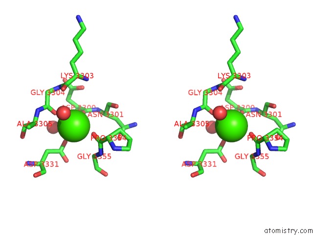
Calcium binding site 1 out of 4 in 1w20
Go back to
Calcium binding site 1 out
of 4 in the Structure of Neuraminidase From English Duck Subtype N6 Complexed with 30 Mm Sialic Acid (Nana, NEU5AC), Crystal Soaked For 3 Hours at 291 K
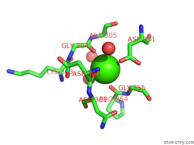
Mono view

Stereo pair view
Mono view
Stereo pair view
A full contact list of Calcium with other atoms in the Ca binding
site number 1 of Structure of Neuraminidase From English Duck Subtype N6 Complexed with 30 Mm Sialic Acid (Nana, NEU5AC), Crystal Soaked For 3 Hours at 291 K within 5.0Å range:
|
Calcium binding site 2 out of 4 in 1w20
Go back to
Calcium binding site 2 out
of 4 in the Structure of Neuraminidase From English Duck Subtype N6 Complexed with 30 Mm Sialic Acid (Nana, NEU5AC), Crystal Soaked For 3 Hours at 291 K
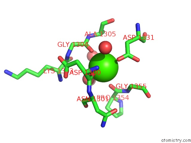
Mono view
Stereo pair view
Mono view
Stereo pair view
A full contact list of Calcium with other atoms in the Ca binding
site number 2 of Structure of Neuraminidase From English Duck Subtype N6 Complexed with 30 Mm Sialic Acid (Nana, NEU5AC), Crystal Soaked For 3 Hours at 291 K within 5.0Å range:
|
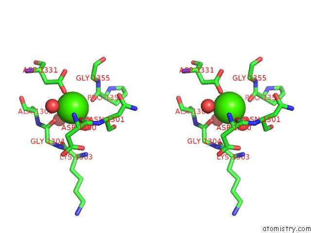
Calcium binding site 3 out of 4 in 1w20
Go back to
Calcium binding site 3 out
of 4 in the Structure of Neuraminidase From English Duck Subtype N6 Complexed with 30 Mm Sialic Acid (Nana, NEU5AC), Crystal Soaked For 3 Hours at 291 K

Mono view

Stereo pair view
Mono view
Stereo pair view
A full contact list of Calcium with other atoms in the Ca binding
site number 3 of Structure of Neuraminidase From English Duck Subtype N6 Complexed with 30 Mm Sialic Acid (Nana, NEU5AC), Crystal Soaked For 3 Hours at 291 K within 5.0Å range:
|
Calcium binding site 4 out of 4 in 1w20
Go back to
Calcium binding site 4 out
of 4 in the Structure of Neuraminidase From English Duck Subtype N6 Complexed with 30 Mm Sialic Acid (Nana, NEU5AC), Crystal Soaked For 3 Hours at 291 K
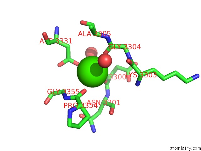
Mono view
Stereo pair view
Mono view
Stereo pair view
A full contact list of Calcium with other atoms in the Ca binding
site number 4 of Structure of Neuraminidase From English Duck Subtype N6 Complexed with 30 Mm Sialic Acid (Nana, NEU5AC), Crystal Soaked For 3 Hours at 291 K within 5.0Å range:
|
Reference:
E.Rudino-Pinera,
P.Tunnah,
S.J.Crennell,
R.G.Webster,
W.G.Laver,
E.F.Garman.
The Crystal Structure of Type A Influenza Virus Neuraminidase of the N6 Subtype Reveals the Existence of Two Separate NEU5AC Binding Sites To Be Published.
Page generated: Tue Jul 8 03:03:42 2025
Last articles
K in 2VQFK in 2VQE
K in 2VWJ
K in 2VXY
K in 2VQW
K in 2VQV
K in 2VQQ
K in 2VQO
K in 2VI5
K in 2VQM