Calcium »
PDB 1vfo-1w7c »
1w52 »
Calcium in PDB 1w52: Crystal Structure of A Proteolyzed Form of Pancreatic Lipase Related Protein 2 From Horse
Protein crystallography data
The structure of Crystal Structure of A Proteolyzed Form of Pancreatic Lipase Related Protein 2 From Horse, PDB code: 1w52
was solved by
J.M.Mancheno,
S.Jayne,
B.Kerfelec,
C.Chapus,
I.Crenon,
J.A.Hermoso,
with X-Ray Crystallography technique. A brief refinement statistics is given in the table below:
| Resolution Low / High (Å) | 26.17 / 2.99 |
| Space group | P 32 2 1 |
| Cell size a, b, c (Å), α, β, γ (°) | 128.426, 128.426, 85.818, 90.00, 90.00, 120.00 |
| R / Rfree (%) | 23.1 / 29.4 |
Calcium Binding Sites:
The binding sites of Calcium atom in the Crystal Structure of A Proteolyzed Form of Pancreatic Lipase Related Protein 2 From Horse
(pdb code 1w52). This binding sites where shown within
5.0 Angstroms radius around Calcium atom.
In total 2 binding sites of Calcium where determined in the Crystal Structure of A Proteolyzed Form of Pancreatic Lipase Related Protein 2 From Horse, PDB code: 1w52:
Jump to Calcium binding site number: 1; 2;
In total 2 binding sites of Calcium where determined in the Crystal Structure of A Proteolyzed Form of Pancreatic Lipase Related Protein 2 From Horse, PDB code: 1w52:
Jump to Calcium binding site number: 1; 2;
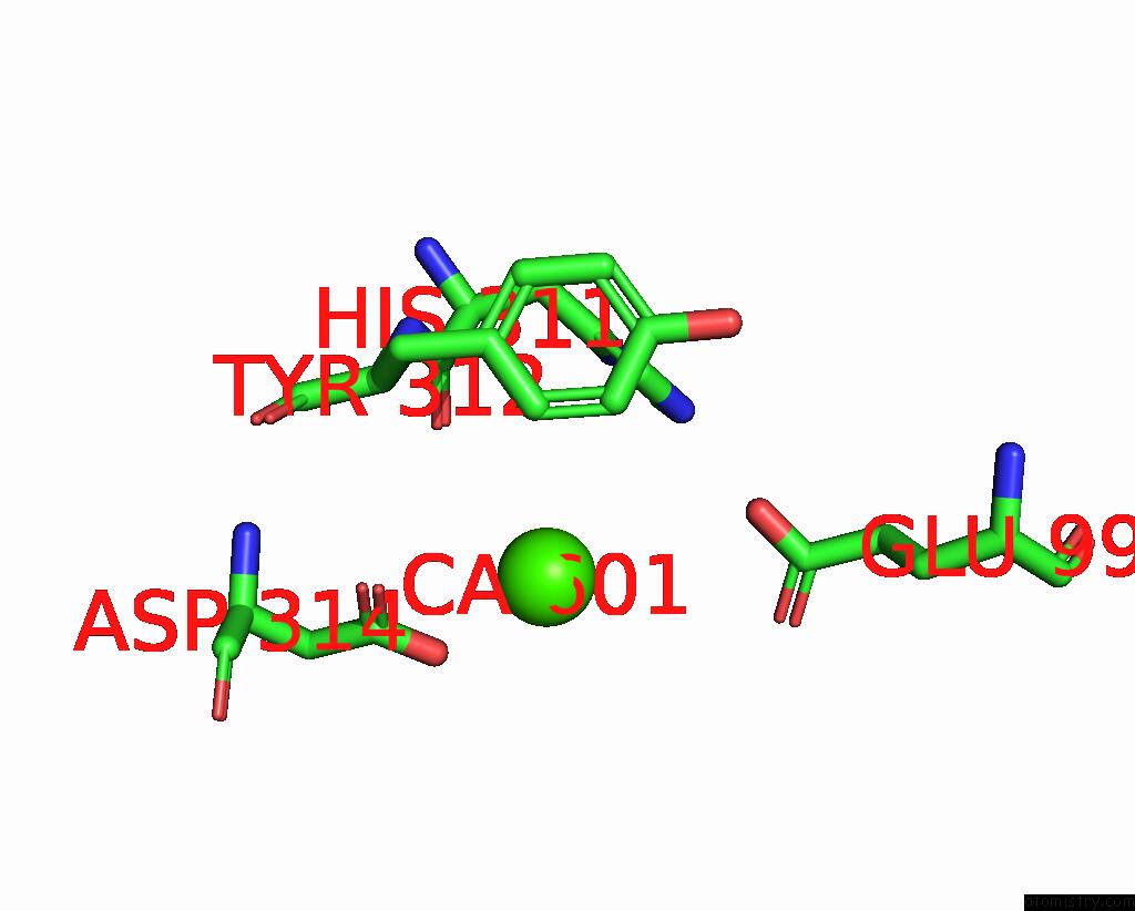
Calcium binding site 1 out of 2 in 1w52
Go back to
Calcium binding site 1 out
of 2 in the Crystal Structure of A Proteolyzed Form of Pancreatic Lipase Related Protein 2 From Horse
Mono view
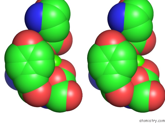
Stereo pair view
Mono view
Stereo pair view
A full contact list of Calcium with other atoms in the Ca binding
site number 1 of Crystal Structure of A Proteolyzed Form of Pancreatic Lipase Related Protein 2 From Horse within 5.0Å range:
|
Calcium binding site 2 out of 2 in 1w52
Go back to
Calcium binding site 2 out
of 2 in the Crystal Structure of A Proteolyzed Form of Pancreatic Lipase Related Protein 2 From Horse
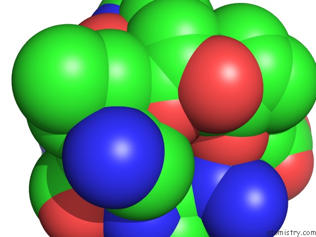
Mono view
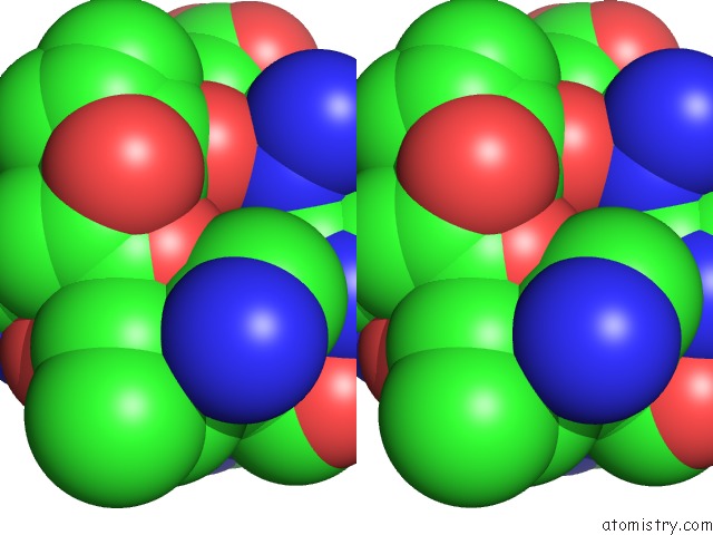
Stereo pair view
Mono view
Stereo pair view
A full contact list of Calcium with other atoms in the Ca binding
site number 2 of Crystal Structure of A Proteolyzed Form of Pancreatic Lipase Related Protein 2 From Horse within 5.0Å range:
|
Reference:
J.M.Mancheno,
S.Jayne,
B.Kerfelec,
C.Chapus,
I.Crenon,
J.A.Hermoso.
Crystalization of A Proteolyzed Form of the Horse Pancreatic Lipase-Related Protein 2: Structural Basis For the Specific Detergent Requirement. Acta Crystallogr.,Sect.D V. 60 2107 2004.
ISSN: ISSN 0907-4449
PubMed: 15502342
DOI: 10.1107/S0907444904024229
Page generated: Tue Jul 8 03:07:56 2025
ISSN: ISSN 0907-4449
PubMed: 15502342
DOI: 10.1107/S0907444904024229
Last articles
K in 4HW8K in 4HYW
K in 4HYV
K in 4HXX
K in 4HX2
K in 4HS7
K in 4H5J
K in 4HLI
K in 4HBW
K in 4H85