Calcium »
PDB 1w7x-1wws »
1wsd »
Calcium in PDB 1wsd: Alkaline M-Protease Form I Crystal Strcuture
Protein crystallography data
The structure of Alkaline M-Protease Form I Crystal Strcuture, PDB code: 1wsd
was solved by
T.Shirai,
A.Suzuki,
T.Yamane,
T.Ashida,
T.Kobayashi,
J.Hitomi,
S.Ito,
with X-Ray Crystallography technique. A brief refinement statistics is given in the table below:
| Resolution Low / High (Å) | 8.00 / 1.50 |
| Space group | P 21 21 21 |
| Cell size a, b, c (Å), α, β, γ (°) | 62.300, 75.500, 47.200, 90.00, 90.00, 90.00 |
| R / Rfree (%) | 17.2 / 19.3 |
Calcium Binding Sites:
The binding sites of Calcium atom in the Alkaline M-Protease Form I Crystal Strcuture
(pdb code 1wsd). This binding sites where shown within
5.0 Angstroms radius around Calcium atom.
In total only one binding site of Calcium was determined in the Alkaline M-Protease Form I Crystal Strcuture, PDB code: 1wsd:
In total only one binding site of Calcium was determined in the Alkaline M-Protease Form I Crystal Strcuture, PDB code: 1wsd:
Calcium binding site 1 out of 1 in 1wsd
Go back to
Calcium binding site 1 out
of 1 in the Alkaline M-Protease Form I Crystal Strcuture
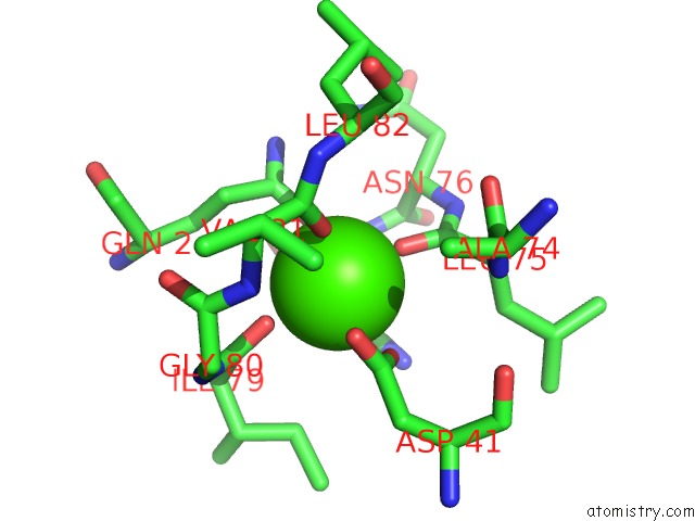
Mono view
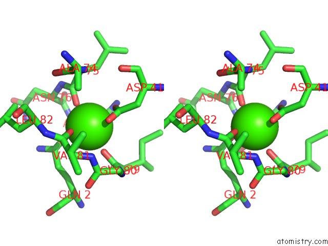
Stereo pair view
Mono view
Stereo pair view
A full contact list of Calcium with other atoms in the Ca binding
site number 1 of Alkaline M-Protease Form I Crystal Strcuture within 5.0Å range:
|
Reference:
T.Shirai,
A.Suzuki,
T.Yamane,
T.Ashida,
T.Kobayashi,
J.Hitomi,
S.Ito.
High-Resolution Crystal Structure of M-Protease: Phylogeny Aided Analysis of the High-Alkaline Adaptation Mechanism Protein Eng. V. 10 627 1997.
ISSN: ISSN 0269-2139
PubMed: 9278275
DOI: 10.1093/PROTEIN/10.6.627
Page generated: Tue Jul 8 03:17:50 2025
ISSN: ISSN 0269-2139
PubMed: 9278275
DOI: 10.1093/PROTEIN/10.6.627
Last articles
Mg in 7AZVMg in 7AYW
Mg in 7AX1
Mg in 7AXY
Mg in 7AUU
Mg in 7AUT
Mg in 7AUR
Mg in 7AUS
Mg in 7AUD
Mg in 7AUP