Calcium »
PDB 1wy9-1xjo »
1wy9 »
Calcium in PDB 1wy9: Crystal Structure of Microglia-Specific Protein, IBA1
Protein crystallography data
The structure of Crystal Structure of Microglia-Specific Protein, IBA1, PDB code: 1wy9
was solved by
M.Yamada,
Y.Imai,
S.Kohsaka,
S.Kamitori,
with X-Ray Crystallography technique. A brief refinement statistics is given in the table below:
| Resolution Low / High (Å) | 35.61 / 2.10 |
| Space group | P 32 2 1 |
| Cell size a, b, c (Å), α, β, γ (°) | 44.056, 44.056, 99.144, 90.00, 90.00, 120.00 |
| R / Rfree (%) | 21.7 / 25.1 |
Calcium Binding Sites:
The binding sites of Calcium atom in the Crystal Structure of Microglia-Specific Protein, IBA1
(pdb code 1wy9). This binding sites where shown within
5.0 Angstroms radius around Calcium atom.
In total only one binding site of Calcium was determined in the Crystal Structure of Microglia-Specific Protein, IBA1, PDB code: 1wy9:
In total only one binding site of Calcium was determined in the Crystal Structure of Microglia-Specific Protein, IBA1, PDB code: 1wy9:
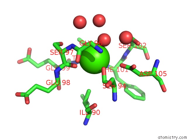
Calcium binding site 1 out of 1 in 1wy9
Go back to
Calcium binding site 1 out
of 1 in the Crystal Structure of Microglia-Specific Protein, IBA1
Mono view

Stereo pair view
Mono view
Stereo pair view
A full contact list of Calcium with other atoms in the Ca binding
site number 1 of Crystal Structure of Microglia-Specific Protein, IBA1 within 5.0Å range:
|
Reference:
M.Yamada,
K.Ohsawa,
Y.Imai,
S.Kohsaka,
S.Kamitori.
X-Ray Structures of the Microglia/Macrophage-Specific Protein IBA1 From Human and Mouse Demonstrate Novel Molecular Conformation Change Induced By Calcium Binding J.Mol.Biol. V. 364 449 2006.
ISSN: ISSN 0022-2836
PubMed: 17011575
DOI: 10.1016/J.JMB.2006.09.027
Page generated: Tue Jul 8 03:22:13 2025
ISSN: ISSN 0022-2836
PubMed: 17011575
DOI: 10.1016/J.JMB.2006.09.027
Last articles
Fe in 2YXOFe in 2YRS
Fe in 2YXC
Fe in 2YNM
Fe in 2YVJ
Fe in 2YP1
Fe in 2YU2
Fe in 2YU1
Fe in 2YQB
Fe in 2YOO