Calcium »
PDB 1wy9-1xjo »
1xhb »
Calcium in PDB 1xhb: The Crystal Structure of Udp-Galnac: Polypeptide Alpha-N- Acetylgalactosaminyltransferase-T1
Enzymatic activity of The Crystal Structure of Udp-Galnac: Polypeptide Alpha-N- Acetylgalactosaminyltransferase-T1
All present enzymatic activity of The Crystal Structure of Udp-Galnac: Polypeptide Alpha-N- Acetylgalactosaminyltransferase-T1:
2.4.1.41;
2.4.1.41;
Protein crystallography data
The structure of The Crystal Structure of Udp-Galnac: Polypeptide Alpha-N- Acetylgalactosaminyltransferase-T1, PDB code: 1xhb
was solved by
T.A.Fritz,
J.H.Hurley,
L.B.Trinh,
J.Shiloach,
L.A.Tabak,
with X-Ray Crystallography technique. A brief refinement statistics is given in the table below:
| Resolution Low / High (Å) | 46.39 / 2.50 |
| Space group | P 43 |
| Cell size a, b, c (Å), α, β, γ (°) | 65.605, 65.605, 125.947, 90.00, 90.00, 90.00 |
| R / Rfree (%) | 21.8 / 25.5 |
Other elements in 1xhb:
The structure of The Crystal Structure of Udp-Galnac: Polypeptide Alpha-N- Acetylgalactosaminyltransferase-T1 also contains other interesting chemical elements:
| Manganese | (Mn) | 1 atom |
Calcium Binding Sites:
The binding sites of Calcium atom in the The Crystal Structure of Udp-Galnac: Polypeptide Alpha-N- Acetylgalactosaminyltransferase-T1
(pdb code 1xhb). This binding sites where shown within
5.0 Angstroms radius around Calcium atom.
In total 2 binding sites of Calcium where determined in the The Crystal Structure of Udp-Galnac: Polypeptide Alpha-N- Acetylgalactosaminyltransferase-T1, PDB code: 1xhb:
Jump to Calcium binding site number: 1; 2;
In total 2 binding sites of Calcium where determined in the The Crystal Structure of Udp-Galnac: Polypeptide Alpha-N- Acetylgalactosaminyltransferase-T1, PDB code: 1xhb:
Jump to Calcium binding site number: 1; 2;
Calcium binding site 1 out of 2 in 1xhb
Go back to
Calcium binding site 1 out
of 2 in the The Crystal Structure of Udp-Galnac: Polypeptide Alpha-N- Acetylgalactosaminyltransferase-T1
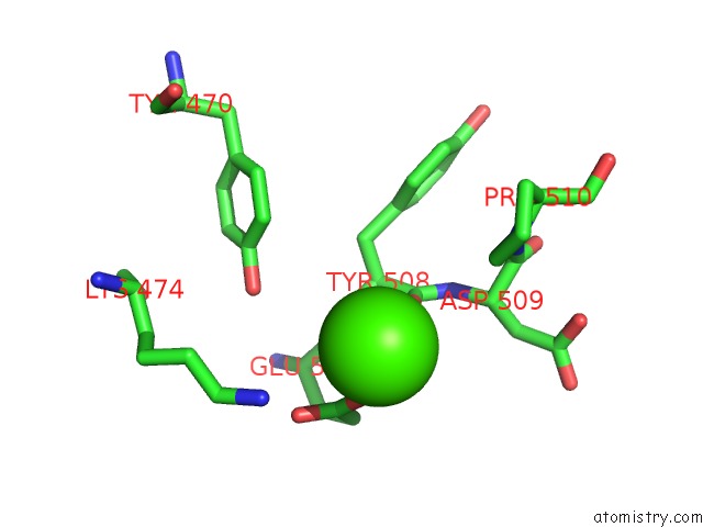
Mono view
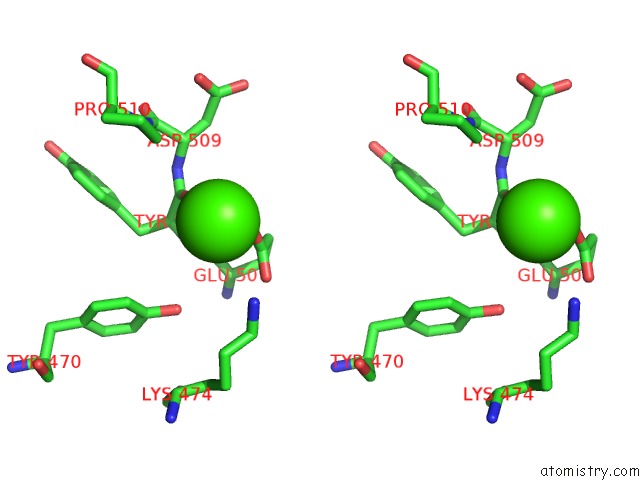
Stereo pair view
Mono view
Stereo pair view
A full contact list of Calcium with other atoms in the Ca binding
site number 1 of The Crystal Structure of Udp-Galnac: Polypeptide Alpha-N- Acetylgalactosaminyltransferase-T1 within 5.0Å range:
|
Calcium binding site 2 out of 2 in 1xhb
Go back to
Calcium binding site 2 out
of 2 in the The Crystal Structure of Udp-Galnac: Polypeptide Alpha-N- Acetylgalactosaminyltransferase-T1
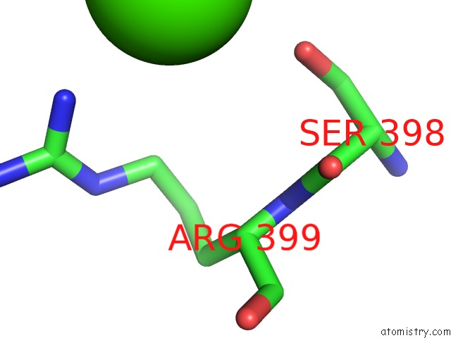
Mono view
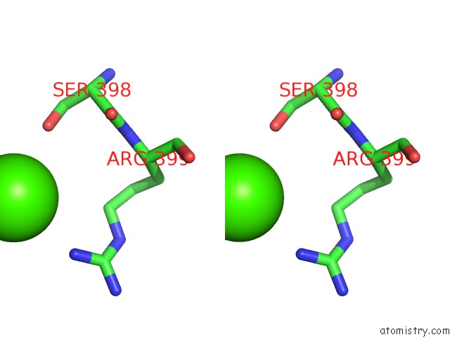
Stereo pair view
Mono view
Stereo pair view
A full contact list of Calcium with other atoms in the Ca binding
site number 2 of The Crystal Structure of Udp-Galnac: Polypeptide Alpha-N- Acetylgalactosaminyltransferase-T1 within 5.0Å range:
|
Reference:
T.A.Fritz,
J.H.Hurley,
L.B.Trinh,
J.Shiloach,
L.A.Tabak.
The Beginnings of Mucin Biosynthesis: the Crystal Structure of Udp-Galnac:Polypeptide {Alpha}-N-Acetylgalactosaminyltransferase-T1 Proc.Natl.Acad.Sci.Usa V. 101 15307 2004.
ISSN: ISSN 0027-8424
PubMed: 15486088
DOI: 10.1073/PNAS.0405657101
Page generated: Tue Jul 8 03:32:06 2025
ISSN: ISSN 0027-8424
PubMed: 15486088
DOI: 10.1073/PNAS.0405657101
Last articles
K in 5FKEK in 5FKD
K in 5FK5
K in 5FJC
K in 5FK3
K in 5FHW
K in 5FCW
K in 5FG0
K in 5FHT
K in 5FG1