Calcium »
PDB 1yvu-1zez »
1z7s »
Calcium in PDB 1z7s: The Crystal Structure of Coxsackievirus A21
Protein crystallography data
The structure of The Crystal Structure of Coxsackievirus A21, PDB code: 1z7s
was solved by
C.Xiao,
C.M.Bator-Kelly,
E.Rieder,
P.R.Chipman,
A.Craig,
R.J.Kuhn,
E.Wimmer,
M.G.Rossmann,
with X-Ray Crystallography technique. A brief refinement statistics is given in the table below:
| Resolution Low / High (Å) | 49.72 / 3.20 |
| Space group | P 42 3 2 1 |
| Cell size a, b, c (Å), α, β, γ (°) | 348.014, 348.014, 348.014, 90.00, 90.00, 90.00 |
| R / Rfree (%) | 22.4 / 23.5 |
Calcium Binding Sites:
The binding sites of Calcium atom in the The Crystal Structure of Coxsackievirus A21
(pdb code 1z7s). This binding sites where shown within
5.0 Angstroms radius around Calcium atom.
In total only one binding site of Calcium was determined in the The Crystal Structure of Coxsackievirus A21, PDB code: 1z7s:
In total only one binding site of Calcium was determined in the The Crystal Structure of Coxsackievirus A21, PDB code: 1z7s:
Calcium binding site 1 out of 1 in 1z7s
Go back to
Calcium binding site 1 out
of 1 in the The Crystal Structure of Coxsackievirus A21
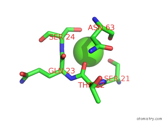
Mono view
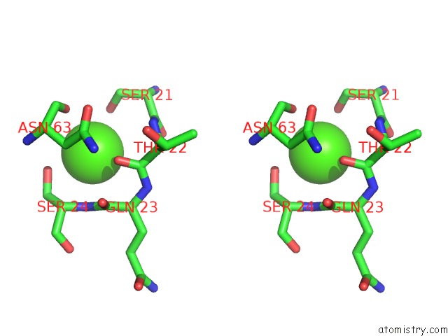
Stereo pair view
Mono view
Stereo pair view
A full contact list of Calcium with other atoms in the Ca binding
site number 1 of The Crystal Structure of Coxsackievirus A21 within 5.0Å range:
|
Reference:
C.Xiao,
C.M.Bator-Kelly,
E.Rieder,
P.R.Chipman,
A.Craig,
R.J.Kuhn,
E.Wimmer,
M.G.Rossmann.
The Crystal Structure of Coxsackievirus A21 and Its Interaction with Icam-1. Structure V. 13 1019 2005.
ISSN: ISSN 0969-2126
PubMed: 16004874
DOI: 10.1016/J.STR.2005.04.011
Page generated: Tue Jul 8 04:02:31 2025
ISSN: ISSN 0969-2126
PubMed: 16004874
DOI: 10.1016/J.STR.2005.04.011
Last articles
Mg in 3AI9Mg in 3AJL
Mg in 3AJK
Mg in 3AHJ
Mg in 3AHI
Mg in 3AHH
Mg in 3AHG
Mg in 3AHF
Mg in 3AHE
Mg in 3AHD