Calcium »
PDB 1zf0-2a2z »
1zot »
Calcium in PDB 1zot: Crystal Structure Analysis of the Cyaa/C-Cam with Pmeapp
Enzymatic activity of Crystal Structure Analysis of the Cyaa/C-Cam with Pmeapp
All present enzymatic activity of Crystal Structure Analysis of the Cyaa/C-Cam with Pmeapp:
4.6.1.1;
4.6.1.1;
Protein crystallography data
The structure of Crystal Structure Analysis of the Cyaa/C-Cam with Pmeapp, PDB code: 1zot
was solved by
Q.Guo,
W.J.Tang,
with X-Ray Crystallography technique. A brief refinement statistics is given in the table below:
| Resolution Low / High (Å) | 34.52 / 2.20 |
| Space group | P 41 21 2 |
| Cell size a, b, c (Å), α, β, γ (°) | 79.674, 79.674, 139.339, 90.00, 90.00, 90.00 |
| R / Rfree (%) | 25.2 / 29.1 |
Other elements in 1zot:
The structure of Crystal Structure Analysis of the Cyaa/C-Cam with Pmeapp also contains other interesting chemical elements:
| Magnesium | (Mg) | 3 atoms |
Calcium Binding Sites:
The binding sites of Calcium atom in the Crystal Structure Analysis of the Cyaa/C-Cam with Pmeapp
(pdb code 1zot). This binding sites where shown within
5.0 Angstroms radius around Calcium atom.
In total 2 binding sites of Calcium where determined in the Crystal Structure Analysis of the Cyaa/C-Cam with Pmeapp, PDB code: 1zot:
Jump to Calcium binding site number: 1; 2;
In total 2 binding sites of Calcium where determined in the Crystal Structure Analysis of the Cyaa/C-Cam with Pmeapp, PDB code: 1zot:
Jump to Calcium binding site number: 1; 2;
Calcium binding site 1 out of 2 in 1zot
Go back to
Calcium binding site 1 out
of 2 in the Crystal Structure Analysis of the Cyaa/C-Cam with Pmeapp

Mono view
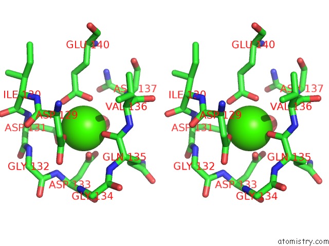
Stereo pair view
Mono view
Stereo pair view
A full contact list of Calcium with other atoms in the Ca binding
site number 1 of Crystal Structure Analysis of the Cyaa/C-Cam with Pmeapp within 5.0Å range:
|
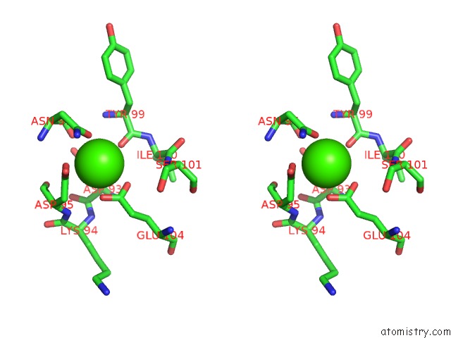
Calcium binding site 2 out of 2 in 1zot
Go back to
Calcium binding site 2 out
of 2 in the Crystal Structure Analysis of the Cyaa/C-Cam with Pmeapp

Mono view
Stereo pair view
Mono view
Stereo pair view
A full contact list of Calcium with other atoms in the Ca binding
site number 2 of Crystal Structure Analysis of the Cyaa/C-Cam with Pmeapp within 5.0Å range:
|
Reference:
Q.Guo,
Y.Shen,
Y.S.Lee,
C.S.Gibbs,
M.Mrksich,
W.J.Tang.
Structural Basis For the Interaction of Bordetella Pertussis Adenylyl Cyclase Toxin with Calmodulin. Embo J. V. 24 3190 2005.
ISSN: ISSN 0261-4189
PubMed: 16138079
DOI: 10.1038/SJ.EMBOJ.7600800
Page generated: Tue Jul 8 04:05:53 2025
ISSN: ISSN 0261-4189
PubMed: 16138079
DOI: 10.1038/SJ.EMBOJ.7600800
Last articles
K in 8OLWK in 8OLJ
K in 8OFD
K in 8OEO
K in 8OED
K in 8OEH
K in 8K1Z
K in 8K1U
K in 8K1V
K in 8K7W