Calcium »
PDB 2a30-2agp »
2a5x »
Calcium in PDB 2a5x: Crystal Structure of A Cross-Linked Actin Dimer
Protein crystallography data
The structure of Crystal Structure of A Cross-Linked Actin Dimer, PDB code: 2a5x
was solved by
D.S.Kudryashov,
M.R.Sawaya,
H.Adisetiyo,
T.Norcross,
G.Hegyi,
E.Reisler,
T.O.Yeates,
with X-Ray Crystallography technique. A brief refinement statistics is given in the table below:
| Resolution Low / High (Å) | 90.00 / 2.49 |
| Space group | C 1 2 1 |
| Cell size a, b, c (Å), α, β, γ (°) | 207.377, 54.372, 36.200, 90.00, 98.62, 90.00 |
| R / Rfree (%) | 19.4 / 25 |
Calcium Binding Sites:
The binding sites of Calcium atom in the Crystal Structure of A Cross-Linked Actin Dimer
(pdb code 2a5x). This binding sites where shown within
5.0 Angstroms radius around Calcium atom.
In total 2 binding sites of Calcium where determined in the Crystal Structure of A Cross-Linked Actin Dimer, PDB code: 2a5x:
Jump to Calcium binding site number: 1; 2;
In total 2 binding sites of Calcium where determined in the Crystal Structure of A Cross-Linked Actin Dimer, PDB code: 2a5x:
Jump to Calcium binding site number: 1; 2;
Calcium binding site 1 out of 2 in 2a5x
Go back to
Calcium binding site 1 out
of 2 in the Crystal Structure of A Cross-Linked Actin Dimer
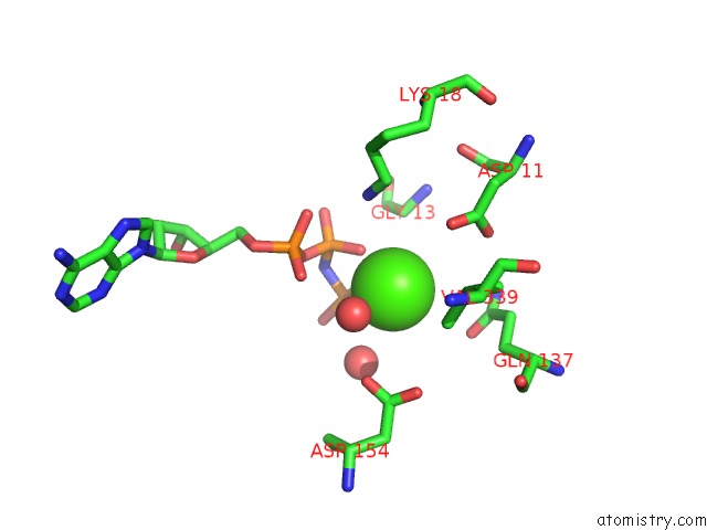
Mono view
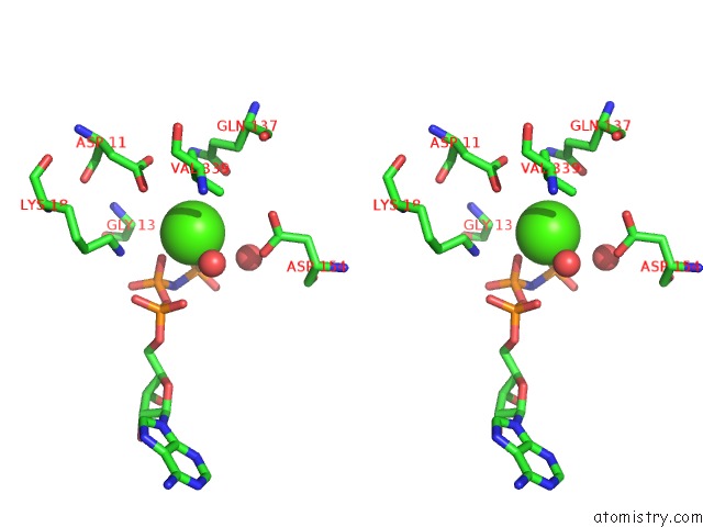
Stereo pair view
Mono view
Stereo pair view
A full contact list of Calcium with other atoms in the Ca binding
site number 1 of Crystal Structure of A Cross-Linked Actin Dimer within 5.0Å range:
|
Calcium binding site 2 out of 2 in 2a5x
Go back to
Calcium binding site 2 out
of 2 in the Crystal Structure of A Cross-Linked Actin Dimer
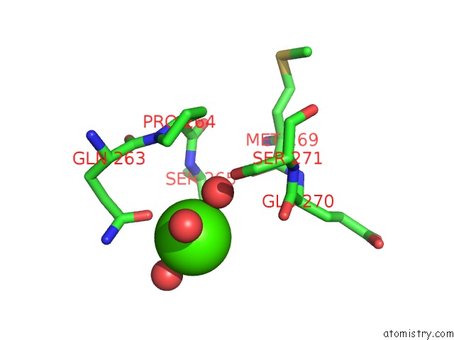
Mono view
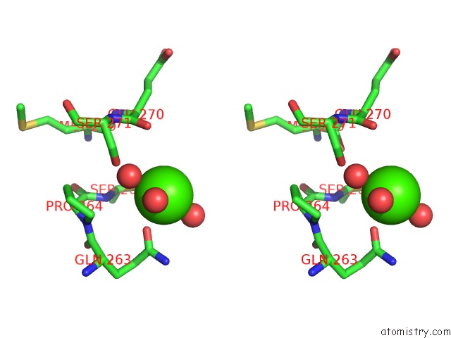
Stereo pair view
Mono view
Stereo pair view
A full contact list of Calcium with other atoms in the Ca binding
site number 2 of Crystal Structure of A Cross-Linked Actin Dimer within 5.0Å range:
|
Reference:
D.S.Kudryashov,
M.R.Sawaya,
H.Adisetiyo,
T.Norcross,
G.Hegyi,
E.Reisler,
T.O.Yeates.
The Crystal Structure of A Cross-Linked Actin Dimer Suggests A Detailed Molecular Interface in F-Actin Proc.Natl.Acad.Sci.Usa V. 102 13105 2005.
ISSN: ISSN 0027-8424
PubMed: 16141336
DOI: 10.1073/PNAS.0506429102
Page generated: Tue Jul 8 04:12:54 2025
ISSN: ISSN 0027-8424
PubMed: 16141336
DOI: 10.1073/PNAS.0506429102
Last articles
Mg in 5DRCMg in 5DRD
Mg in 5DQL
Mg in 5DR2
Mg in 5DQZ
Mg in 5DQK
Mg in 5DOU
Mg in 5DQH
Mg in 5DQG
Mg in 5DPH